5KTG

Crystal structure of mouse Bak BH3-in-groove homodimer (GFP)


Experimental Data Snapshot

  • Method: X-RAY DIFFRACTION
  • Resolution: 2.80 Å
  • R-Value Free: 0.275 
  • R-Value Work: 0.244 
  • R-Value Observed: 0.245 

wwPDB Validation   3D Report Full Report


This is version 1.4 of the entry. See complete history


Literature

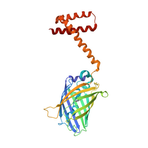
Assembly of Bak homodimers into higher order homooligomers in the mitochondrial apoptotic pore.

Mandal, T.Shin, S.Aluvila, S.Chen, H.C.Grieve, C.Choe, J.Y.Cheng, E.H.Hustedt, E.J.Oh, K.J.

(2016) Sci Rep 6: 30763-30763

  • DOI: https://doi.org/10.1038/srep30763
  • Primary Citation of Related Structures:  
    5KTG

  • PubMed Abstract: 

    In mitochondrial apoptosis, Bak is activated by death signals to form pores of unknown structure on the mitochondrial outer membrane via homooligomerization. Cytochrome c and other apoptotic factors are released from the intermembrane space through these pores, initiating downstream apoptosis events. Using chemical crosslinking and double electron electron resonance (DEER)-derived distance measurements between specific structural elements in Bak, here we clarify how the Bak pore is assembled. We propose that previously described BH3-in-groove homodimers (BGH) are juxtaposed via the 'α3/α5' interface, in which the C-termini of helices α3 and α5 are in close proximity between two neighboring Bak homodimers. This interface is observed concomitantly with the well-known 'α6:α6' interface. We also mapped the contacts between Bak homodimers and the lipid bilayer based on EPR spectroscopy topology studies. Our results suggest a model for the lipidic Bak pore, whereby the mitochondrial targeting C-terminal helix does not change topology to accommodate the lining of the pore lumen by BGH.


  • Organizational Affiliation

    Department of Biochemistry and Molecular Biology, Rosalind Franklin University of Medicine and Science, North Chicago, Illinois 60064, USA.


Macromolecules
Find similar proteins by:  (by identity cutoff)  |  3D Structure
Entity ID: 1
MoleculeChains Sequence LengthOrganismDetailsImage
Green fluorescent protein, Bcl-2 homologous antagonist/killer
A, B
309Aequorea victoriaMus musculus
This entity is chimeric
Mutation(s): 1 
Gene Names: GFPBak1Bak
UniProt
Find proteins for P42212 (Aequorea victoria)
Explore P42212 
Go to UniProtKB:  P42212
Find proteins for O08734 (Mus musculus)
Explore O08734 
Go to UniProtKB:  O08734
Entity Groups  
Sequence Clusters30% Identity50% Identity70% Identity90% Identity95% Identity100% Identity
UniProt GroupsP42212O08734
Sequence Annotations
Expand
  • Reference Sequence
Small Molecules
Modified Residues  1 Unique
IDChains TypeFormula2D DiagramParent
CR2
Query on CR2
A, B
L-PEPTIDE LINKINGC13 H13 N3 O4GLY, TYR, GLY
Experimental Data & Validation

Experimental Data

  • Method: X-RAY DIFFRACTION
  • Resolution: 2.80 Å
  • R-Value Free: 0.275 
  • R-Value Work: 0.244 
  • R-Value Observed: 0.245 
  • Space Group: P 31 2 1
Unit Cell:
Length ( Å )Angle ( ˚ )
a = 171.641α = 90
b = 171.641β = 90
c = 98.194γ = 120
Software Package:
Software NamePurpose
PHENIXrefinement
HKL-3000data reduction
SCALEPACKdata scaling
PHASERphasing

Structure Validation

View Full Validation Report


Entry History & Funding Information

Deposition Data


Funding OrganizationLocationGrant Number
National Institutes of Health/National Institute of General Medical Sciences (NIH/NIGMS)United States5 R01 GM097508

Revision History  (Full details and data files)

  • Version 1.0: 2016-08-17
    Type: Initial release
  • Version 1.1: 2017-09-13
    Changes: Author supporting evidence, Derived calculations
  • Version 1.2: 2019-12-25
    Changes: Author supporting evidence
  • Version 1.3: 2023-10-04
    Changes: Data collection, Database references, Derived calculations, Refinement description
  • Version 1.4: 2023-11-15
    Changes: Data collection