5KQP

Crystal structure of Apo-form LMW-PTP


Experimental Data Snapshot

  • Method: X-RAY DIFFRACTION
  • Resolution: 2.05 Å
  • R-Value Free: 0.266 
  • R-Value Work: 0.193 
  • R-Value Observed: 0.196 

wwPDB Validation   3D Report Full Report


This is version 1.1 of the entry. See complete history


Literature

Inhibition of low molecular weight protein tyrosine phosphatase by an induced-fit mechanism.

He, R.Wang, J.Yu, Z.H.Zhang, R.Y.Liu, S.Wu, L.Zhang, Z.Y.

(2016) J Med Chem 

  • DOI: https://doi.org/10.1021/acs.jmedchem.6b00993
  • Primary Citation of Related Structures:  
    5KQG, 5KQL, 5KQM, 5KQP

  • PubMed Abstract: 

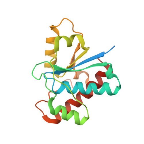
    The low molecular weight protein tyrosine phosphatase (LMW-PTP) is a regulator of a number of signaling pathways and has been implicated as a potential target for oncology and diabetes/obesity. There is significant therapeutic interest in developing potent and selective inhibitors to control LMW-PTP activity. We report the discovery of a novel class of LMW-PTP inhibitors derived from sulfophenyl acetic amide (SPAA), some of which exhibit greater than 50-fold preference for LMW-PTP over a large panel of PTPs. X-ray crystallography reveals that binding of SPAA-based inhibitors induces a striking conformational change in the LMW-PTP active site, leading to the formation of a previously undisclosed hydrophobic pocket to accommodate the α-phenyl ring in the ligand. This induced-fit mechanism is likely a major contributor responsible for the exquisite inhibitor selectivity.


  • Organizational Affiliation

    Department of Medicinal Chemistry and Molecular Pharmacology, Department of Chemistry, Center for Cancer Research, and Institute for Drug Discovery, Purdue University , 720 Clinic Drive, West Lafayette, Indiana 47907, United States.


Macromolecules
Find similar proteins by:  (by identity cutoff)  |  3D Structure
Entity ID: 1
MoleculeChains Sequence LengthOrganismDetailsImage
Low molecular weight phosphotyrosine protein phosphatase178Homo sapiensMutation(s): 0 
Gene Names: ACP1
EC: 3.1.3.48 (PDB Primary Data), 3.1.3.2 (PDB Primary Data)
UniProt & NIH Common Fund Data Resources
Find proteins for P24666 (Homo sapiens)
Explore P24666 
Go to UniProtKB:  P24666
PHAROS:  P24666
GTEx:  ENSG00000143727 
Entity Groups  
Sequence Clusters30% Identity50% Identity70% Identity90% Identity95% Identity100% Identity
UniProt GroupP24666
Sequence Annotations
Expand
  • Reference Sequence
Experimental Data & Validation

Experimental Data

  • Method: X-RAY DIFFRACTION
  • Resolution: 2.05 Å
  • R-Value Free: 0.266 
  • R-Value Work: 0.193 
  • R-Value Observed: 0.196 
  • Space Group: P 21 21 21
Unit Cell:
Length ( Å )Angle ( ˚ )
a = 31.957α = 90
b = 55.453β = 90
c = 96.326γ = 90
Software Package:
Software NamePurpose
PHENIXrefinement
HKL-3000data reduction
HKL-3000data scaling
MOLREPphasing

Structure Validation

View Full Validation Report


Entry History 

Deposition Data

Revision History  (Full details and data files)

  • Version 1.0: 2016-10-12
    Type: Initial release
  • Version 1.1: 2023-10-04
    Changes: Data collection, Database references, Derived calculations, Refinement description