5KGJ

X-ray structure of a glucosamine N-Acetyltransferase from Clostridium acetobutylicum in complex with galactosamine


Experimental Data Snapshot

  • Method: X-RAY DIFFRACTION
  • Resolution: 1.90 Å
  • R-Value Free: 0.215 
  • R-Value Work: 0.167 
  • R-Value Observed: 0.170 

wwPDB Validation   3D Report Full Report


Ligand Structure Quality Assessment 


This is version 2.1 of the entry. See complete history


Literature

Structural Studies on a Glucosamine/Glucosaminide N-Acetyltransferase.

Dopkins, B.J.Tipton, P.A.Thoden, J.B.Holden, H.M.

(2016) Biochemistry 55: 4495-4508

  • DOI: https://doi.org/10.1021/acs.biochem.6b00536
  • Primary Citation of Related Structures:  
    5KF1, 5KF2, 5KF8, 5KF9, 5KGA, 5KGH, 5KGJ, 5KGP

  • PubMed Abstract: 

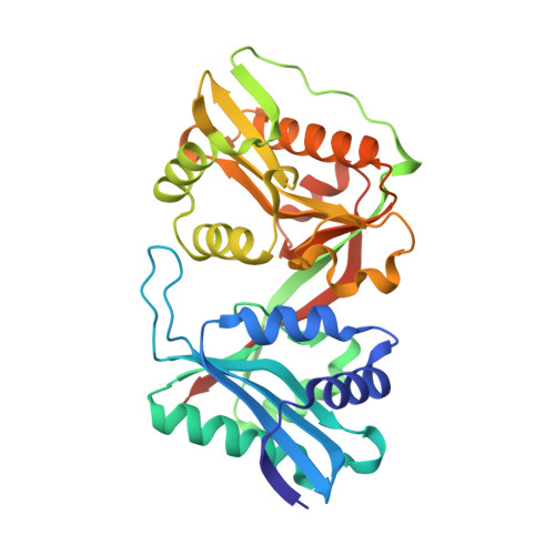
    Glucosamine/glucosaminide N-acetyltransferase or GlmA catalyzes the transfer of an acetyl group from acetyl CoA to the primary amino group of glucosamine. The enzyme from Clostridium acetobutylicum is thought to be involved in cell wall rescue. In addition to glucosamine, GlmA has been shown to function on di- and trisaccharides of glucosamine as well. Here we present a structural and kinetic analysis of the enzyme. For this investigation, eight structures were determined to resolutions of 2.0 Å or better. The overall three-dimensional fold of GlmA places it into the tandem GNAT superfamily. Each subunit of the dimer folds into two distinct domains which exhibit high three-dimensional structural similarity. Whereas both domains bind acetyl CoA, it is the C-terminal domain that is catalytically competent. On the basis of the various structures determined in this investigation, two amino acid residues were targeted for further study: Asp 287 and Tyr 297. Although their positions in the active site suggested that they may play key roles in catalysis by functioning as active site bases and acids, respectively, this was not borne out by characterization of the D287N and Y297F variants. The kinetic properties revealed that both residues were important for substrate binding but had no critical roles as acid/base catalysts. Kinetic analyses also indicated that GlmA follows an ordered mechanism with acetyl CoA binding first followed by glucosamine. The product N-acetylglucosamine is then released prior to CoA. The investigation described herein provides significantly new information on enzymes belonging to the tandem GNAT superfamily.


  • Organizational Affiliation

    Department of Biochemistry, University of Wisconsin , Madison, Wisconsin 53706, United States.


Macromolecules
Find similar proteins by:  (by identity cutoff)  |  3D Structure
Entity ID: 1
MoleculeChains Sequence LengthOrganismDetailsImage
Predicted acetyltransferase328Clostridium acetobutylicum ATCC 824Mutation(s): 0 
Gene Names: CA_C0184
UniProt
Find proteins for Q97ML2 (Clostridium acetobutylicum (strain ATCC 824 / DSM 792 / JCM 1419 / LMG 5710 / VKM B-1787))
Explore Q97ML2 
Go to UniProtKB:  Q97ML2
Entity Groups  
Sequence Clusters30% Identity50% Identity70% Identity90% Identity95% Identity100% Identity
UniProt GroupQ97ML2
Sequence Annotations
Expand
  • Reference Sequence
Small Molecules
Ligands 5 Unique
IDChains Name / Formula / InChI Key2D Diagram3D Interactions
ACO
Query on ACO

Download Ideal Coordinates CCD File 
D [auth A]ACETYL COENZYME *A
C23 H38 N7 O17 P3 S
ZSLZBFCDCINBPY-ZSJPKINUSA-N
COA
Query on COA

Download Ideal Coordinates CCD File 
B [auth A]COENZYME A
C21 H36 N7 O16 P3 S
RGJOEKWQDUBAIZ-IBOSZNHHSA-N
EP1
Query on EP1

Download Ideal Coordinates CCD File 
H [auth A]3-[4-(2-HYDROXYETHYL)PIPERAZIN-1-YL]PROPANE-1-SULFONIC ACID
C9 H20 N2 O4 S
OWXMKDGYPWMGEB-UHFFFAOYSA-N
X6X
Query on X6X

Download Ideal Coordinates CCD File 
C [auth A]2-amino-2-deoxy-alpha-D-galactopyranose
C6 H13 N O5
MSWZFWKMSRAUBD-DVKNGEFBSA-N
EDO
Query on EDO

Download Ideal Coordinates CCD File 
E [auth A],
F [auth A],
G [auth A]
1,2-ETHANEDIOL
C2 H6 O2
LYCAIKOWRPUZTN-UHFFFAOYSA-N
Binding Affinity Annotations 
IDSourceBinding Affinity
ACO Binding MOAD:  5KGJ Ki: 3000 (nM) from 1 assay(s)
Experimental Data & Validation

Experimental Data

  • Method: X-RAY DIFFRACTION
  • Resolution: 1.90 Å
  • R-Value Free: 0.215 
  • R-Value Work: 0.167 
  • R-Value Observed: 0.170 
  • Space Group: C 1 2 1
Unit Cell:
Length ( Å )Angle ( ˚ )
a = 119.076α = 90
b = 43.84β = 120.81
c = 74.169γ = 90
Software Package:
Software NamePurpose
REFMACrefinement
SAINTdata reduction
SADABSdata scaling
PHASERphasing

Structure Validation

View Full Validation Report


Ligand Structure Quality Assessment 


Entry History & Funding Information

Deposition Data


Funding OrganizationLocationGrant Number
National Institutes of Health/National Institute of General Medical Sciences (NIH/NIGMS)United StatesGM115921

Revision History  (Full details and data files)

  • Version 1.0: 2016-07-06
    Type: Initial release
  • Version 1.1: 2016-07-13
    Changes: Database references
  • Version 1.2: 2016-08-24
    Changes: Database references
  • Version 1.3: 2017-09-27
    Changes: Author supporting evidence, Database references, Derived calculations
  • Version 1.4: 2019-12-25
    Changes: Author supporting evidence
  • Version 2.0: 2020-07-29
    Type: Remediation
    Reason: Carbohydrate remediation
    Changes: Atomic model, Data collection, Derived calculations
  • Version 2.1: 2023-09-27
    Changes: Data collection, Database references, Refinement description, Structure summary