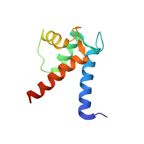
5K89

Crystal Structure of Human Calcium-Bound S100A1


Experimental Data Snapshot

  • Method: X-RAY DIFFRACTION
  • Resolution: 2.25 Å
  • R-Value Free: 0.290 
  • R-Value Work: 0.275 
  • R-Value Observed: 0.275 

wwPDB Validation   3D Report Full Report


This is version 1.4 of the entry. See complete history


Literature

X-ray crystal structure of human calcium-bound S100A1.

Melville, Z.Aligholizadeh, E.McKnight, L.E.Weber, D.J.Pozharski, E.Weber, D.J.

(2017) Acta Crystallogr F Struct Biol Commun 73: 215-221

  • DOI: https://doi.org/10.1107/S2053230X17003983
  • Primary Citation of Related Structures:  
    5K89

  • PubMed Abstract: 

    S100A1 is a member of the S100 family of Ca 2+ -binding proteins and regulates several cellular processes, including those involved in Ca 2+ signaling and cardiac and skeletal muscle function. In Alzheimer's disease, brain S100A1 is overexpressed and gives rise to disease pathologies, making it a potential therapeutic target. The 2.25 Å resolution crystal structure of Ca 2+ -S100A1 is solved here and is compared with the structures of other S100 proteins, most notably S100B, which is a highly homologous S100-family member that is implicated in the progression of malignant melanoma. The observed structural differences in S100A1 versus S100B provide insights regarding target protein-binding specificity and for targeting these two S100 proteins in human diseases using structure-based drug-design approaches.


  • Organizational Affiliation

    Center for Biomolecular Therapeutics, Department of Biochemistry and Molecular Biology, University of Maryland Baltimore, 108 North Greene Street, Baltimore, MD 21201, USA.


Macromolecules
Find similar proteins by:  (by identity cutoff)  |  3D Structure
Entity ID: 1
MoleculeChains Sequence LengthOrganismDetailsImage
Protein S100-A1A,
B [auth C],
C [auth H]
94Homo sapiensMutation(s): 0 
Gene Names: S100A1S100A
UniProt & NIH Common Fund Data Resources
Find proteins for P23297 (Homo sapiens)
Explore P23297 
Go to UniProtKB:  P23297
PHAROS:  P23297
GTEx:  ENSG00000160678 
Entity Groups  
Sequence Clusters30% Identity50% Identity70% Identity90% Identity95% Identity100% Identity
UniProt GroupP23297
Sequence Annotations
Expand
  • Reference Sequence
Experimental Data & Validation

Experimental Data

  • Method: X-RAY DIFFRACTION
  • Resolution: 2.25 Å
  • R-Value Free: 0.290 
  • R-Value Work: 0.275 
  • R-Value Observed: 0.275 
  • Space Group: P 32 2 1
Unit Cell:
Length ( Å )Angle ( ˚ )
a = 53.547α = 90
b = 53.547β = 90
c = 193.387γ = 120
Software Package:
Software NamePurpose
PHENIXrefinement
Blu-Icedata collection
PDB_EXTRACTdata extraction
autoXDSdata reduction
SCALAdata scaling
Cootmodel building
BALBESphasing

Structure Validation

View Full Validation Report


Entry History & Funding Information

Deposition Data


Funding OrganizationLocationGrant Number
National Institutes of Health/National Cancer Institute (NIH/NCI)United StatesCA-107331
National Institutes of Health/National Institute of General Medical Sciences (NIH/NIGMS)United StatesGM-58888
National Institutes of Health/National Institute of Arthritis and Musculoskeletal and Skin Diseases (NIH/NIAMS)United StatesAR007592

Revision History  (Full details and data files)

  • Version 1.0: 2017-04-12
    Type: Initial release
  • Version 1.1: 2017-09-13
    Changes: Author supporting evidence, Derived calculations
  • Version 1.2: 2018-05-16
    Changes: Data collection, Database references
  • Version 1.3: 2019-12-04
    Changes: Author supporting evidence
  • Version 1.4: 2023-09-27
    Changes: Data collection, Database references, Derived calculations, Refinement description