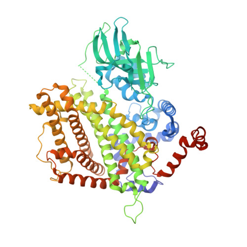
5K3I

Crystal structure of Acyl-CoA oxidase-1 in Caenorhabditis elegans complexed with FAD and ATP

  • Classification: OXIDOREDUCTASE
  • Organism(s): Caenorhabditis elegans
  • Expression System: Escherichia coli
  • Mutation(s): Yes 

  • Deposited: 2016-05-19 Released: 2016-08-24 
  • Deposition Author(s): Zhang, X., Li, K., Jones, R.A., Bruner, S.D., Butcher, R.A.
  • Funding Organization(s): Research Corporation for Science Advancement, Ellison Medical Foundation, National Science Foundation (NSF, United States), National Institutes of Health/National Institute of General Medical Sciences (NIH/NIGMS), Alfred P. Sloan Foundation

Experimental Data Snapshot

  • Method: X-RAY DIFFRACTION
  • Resolution: 2.68 Å
  • R-Value Free: 0.232 
  • R-Value Work: 0.217 
  • R-Value Observed: 0.218 

wwPDB Validation   3D Report Full Report


Ligand Structure Quality Assessment 


This is version 1.6 of the entry. See complete history


Literature

Structural characterization of acyl-CoA oxidases reveals a direct link between pheromone biosynthesis and metabolic state in Caenorhabditis elegans.

Zhang, X.Li, K.Jones, R.A.Bruner, S.D.Butcher, R.A.

(2016) Proc Natl Acad Sci U S A 113: 10055-10060

  • DOI: https://doi.org/10.1073/pnas.1608262113
  • Primary Citation of Related Structures:  
    5K3G, 5K3H, 5K3I, 5K3J

  • PubMed Abstract: 

    Caenorhabditis elegans secretes ascarosides as pheromones to communicate with other worms and to coordinate the development and behavior of the population. Peroxisomal β-oxidation cycles shorten the side chains of ascaroside precursors to produce the short-chain ascaroside pheromones. Acyl-CoA oxidases, which catalyze the first step in these β-oxidation cycles, have different side chain-length specificities and enable C. elegans to regulate the production of specific ascaroside pheromones. Here, we determine the crystal structure of the acyl-CoA oxidase 1 (ACOX-1) homodimer and the ACOX-2 homodimer bound to its substrate. Our results provide a molecular basis for the substrate specificities of the acyl-CoA oxidases and reveal why some of these enzymes have a very broad substrate range, whereas others are quite specific. Our results also enable predictions to be made for the roles of uncharacterized acyl-CoA oxidases in C. elegans and in other nematode species. Remarkably, we show that most of the C. elegans acyl-CoA oxidases that participate in ascaroside biosynthesis contain a conserved ATP-binding pocket that lies at the dimer interface, and we identify key residues in this binding pocket. ATP binding induces a structural change that is associated with tighter binding of the FAD cofactor. Mutations that disrupt ATP binding reduce FAD binding and reduce enzyme activity. Thus, ATP may serve as a regulator of acyl-CoA oxidase activity, thereby directly linking ascaroside biosynthesis to ATP concentration and metabolic state.


  • Organizational Affiliation

    Department of Chemistry, University of Florida, Gainesville, FL 32611.


Macromolecules
Find similar proteins by:  (by identity cutoff)  |  3D Structure
Entity ID: 1
MoleculeChains Sequence LengthOrganismDetailsImage
Acyl-coenzyme A oxidase
A, B, C, D, E
A, B, C, D, E, F, G, H
684Caenorhabditis elegansMutation(s): 1 
Gene Names: acox-1CELE_F08A8.1F08A8.1
UniProt
Find proteins for O62140 (Caenorhabditis elegans)
Explore O62140 
Go to UniProtKB:  O62140
Entity Groups  
Sequence Clusters30% Identity50% Identity70% Identity90% Identity95% Identity100% Identity
UniProt GroupO62140
Sequence Annotations
Expand
  • Reference Sequence
Small Molecules
Ligands 3 Unique
IDChains Name / Formula / InChI Key2D Diagram3D Interactions
FAD
Query on FAD

Download Ideal Coordinates CCD File 
AA [auth G]
DA [auth H]
I [auth A]
M [auth B]
O [auth C]
AA [auth G],
DA [auth H],
I [auth A],
M [auth B],
O [auth C],
T [auth D],
U [auth E],
X [auth F]
FLAVIN-ADENINE DINUCLEOTIDE
C27 H33 N9 O15 P2
VWWQXMAJTJZDQX-UYBVJOGSSA-N
ATP
Query on ATP

Download Ideal Coordinates CCD File 
CA [auth G]
EA [auth H]
J [auth A]
N [auth B]
Q [auth C]
CA [auth G],
EA [auth H],
J [auth A],
N [auth B],
Q [auth C],
R [auth D],
W [auth E],
Y [auth F]
ADENOSINE-5'-TRIPHOSPHATE
C10 H16 N5 O13 P3
ZKHQWZAMYRWXGA-KQYNXXCUSA-N
MG
Query on MG

Download Ideal Coordinates CCD File 
BA [auth G]
FA [auth H]
K [auth A]
L [auth B]
P [auth C]
BA [auth G],
FA [auth H],
K [auth A],
L [auth B],
P [auth C],
S [auth D],
V [auth E],
Z [auth F]
MAGNESIUM ION
Mg
JLVVSXFLKOJNIY-UHFFFAOYSA-N
Experimental Data & Validation

Experimental Data

  • Method: X-RAY DIFFRACTION
  • Resolution: 2.68 Å
  • R-Value Free: 0.232 
  • R-Value Work: 0.217 
  • R-Value Observed: 0.218 
  • Space Group: P 1 21 1
Unit Cell:
Length ( Å )Angle ( ˚ )
a = 152.339α = 90
b = 141.501β = 116.76
c = 154.968γ = 90
Software Package:
Software NamePurpose
PHENIXrefinement
XDSdata reduction
Aimlessdata scaling
PHASERphasing
PHENIXmodel building

Structure Validation

View Full Validation Report


Ligand Structure Quality Assessment 


Entry History & Funding Information

Deposition Data


Funding OrganizationLocationGrant Number
Research Corporation for Science AdvancementUnited States22844
Ellison Medical FoundationUnited StatesAG-NS-0963-12
National Science Foundation (NSF, United States)United States1555050
National Institutes of Health/National Institute of General Medical Sciences (NIH/NIGMS)United StatesGM087533
Alfred P. Sloan FoundationUnited StatesBR2014-071

Revision History  (Full details and data files)

  • Version 1.0: 2016-08-24
    Type: Initial release
  • Version 1.1: 2016-09-07
    Changes: Database references
  • Version 1.2: 2016-09-14
    Changes: Database references
  • Version 1.3: 2017-09-27
    Changes: Author supporting evidence, Database references, Derived calculations
  • Version 1.4: 2019-11-27
    Changes: Author supporting evidence
  • Version 1.5: 2023-09-27
    Changes: Data collection, Database references, Derived calculations, Refinement description
  • Version 1.6: 2024-01-31
    Changes: Refinement description