Chitin oligosaccharide binding to the lysin motif of a novel type of chitinase from the multicellular green alga, Volvox carteri.
Kitaoku, Y., Fukamizo, T., Numata, T., Ohnuma, T.(2017) Plant Mol Biol 93: 97-108
- PubMed: 27807643
- DOI: https://doi.org/10.1007/s11103-016-0549-5
- Primary Citation of Related Structures:
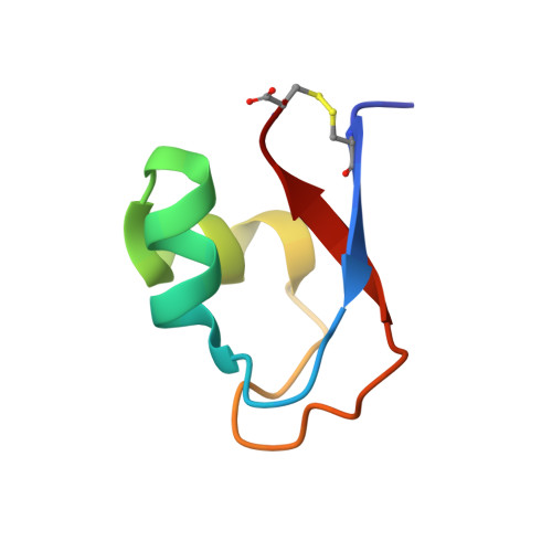
5K2L - PubMed Abstract:
The chitinase-mediated defense system in higher plants has been intensively studied from physiological and structural viewpoints. However, the defense system in the most primitive plant species, such as green algae, has not yet been elucidated in details. In this study, we solved the crystal structure of a family CBM-50 LysM module attached to the N-terminus of chitinase from Volvox carteri, and successfully analyzed its chitin-binding ability by NMR spectroscopy and isothermal titration calorimetry. Trp96 of the LysM module appeared to make a CH-π stacking interaction with the reducing end sugar residue of the ligand. We believe the data included in this manuscript provide novel insights into the molecular basis of chitinase-mediated defense system in green algae. A chitinase from the multicellular green alga, Volvox carteri, contains two N-terminal lysin motifs (VcLysM1 and VcLysM2), that belong to the CBM-50 family, in addition to a catalytic domain. We produced a recombinant protein of VcLysM2 in order to examine its structure and function. The X-ray crystal structure of VcLysM2 was successfully solved at a resolution of 1.2 Å, and revealed that the protein adopts the βααβ fold typical of members belonging to the CBM-50 family. NMR spectra of 13 C- and 15 N-labeled proteins were analyzed in order to completely assign the main chain resonances of the 1 H, 15 N-HSQC spectrum in a sequential manner. NMR-based titration experiments of chitin oligosaccharides, (GlcNAc) n (n = 3-6), revealed the ligand-binding site of VcLysM2, in which the Trp96 side chain appeared to interact with the terminal GlcNAc residue of the ligand. We then mutated Trp96 to alanine (VcLysM2-W96A), and the mutant protein was characterized. Based on isothermal titration calorimetry, the affinity of (GlcNAc) 6 toward VcLysM2 (-6.9 kcal/mol) was found to be markedly higher than that of (GlcNAc) 3 (-4.1 kcal/mol), whereas the difference in affinities between (GlcNAc) 6 and (GlcNAc) 3 in VcLysM2-W96A (-5.1 and -4.0 kcal/mol, respectively) was only moderate. This suggests that the Trp96 side chain of VcLysM2 interacts with the sugar residue of (GlcNAc) 6 not with (GlcNAc) 3 . VcLysM2 appears to preferentially bind (GlcNAc) n with longer chains and plays a major role in the degradation of the chitinous components of enzyme targets.
Organizational Affiliation:
Department of Advanced Bioscience, Kindai University, 3327-204 Nakamachi, Nara, 631-8505, Japan.