5JQP

Crystal structure of ER glucosidase II heterodimeric complex consisting of catalytic subunit and the binding domain of regulatory subunit


Experimental Data Snapshot

  • Method: X-RAY DIFFRACTION
  • Resolution: 2.20 Å
  • R-Value Free: 0.206 
  • R-Value Work: 0.153 
  • R-Value Observed: 0.155 

wwPDB Validation   3D Report Full Report


This is version 1.3 of the entry. See complete history


Literature

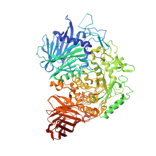
Interaction mode between catalytic and regulatory subunits in glucosidase II involved in ER glycoprotein quality control.

Satoh, T.Toshimori, T.Noda, M.Uchiyama, S.Kato, K.

(2016) Protein Sci 25: 2095-2101

  • DOI: https://doi.org/10.1002/pro.3031
  • Primary Citation of Related Structures:  
    5JQP

  • PubMed Abstract: 

    The glycoside hydrolase family 31 (GH31) α-glucosidases play vital roles in catabolic and regulated degradation, including the α-subunit of glucosidase II (GIIα), which catalyzes trimming of the terminal glucose residues of N-glycan in glycoprotein processing coupled with quality control in the endoplasmic reticulum (ER). Among the known GH31 enzymes, only GIIα functions with its binding partner, regulatory β-subunit (GIIβ), which harbors a lectin domain for substrate recognition. Although the structural data have been reported for GIIα and the GIIβ lectin domain, the interaction mode between GIIα and GIIβ remains unknown. Here, we determined the structure of a complex formed between GIIα and the GIIα-binding domain of GIIβ, thereby providing a structural basis underlying the functional extension of this unique GH31 enzyme.


  • Organizational Affiliation

    Graduate School of Pharmaceutical Sciences, Nagoya City University, 3-1 Tanabe-dori, Mizuho-ku, Nagoya, 467-8603, Japan. tadashisatoh@phar.nagoya-cu.ac.jp.


Macromolecules
Find similar proteins by:  (by identity cutoff)  |  3D Structure
Entity ID: 1
MoleculeChains Sequence LengthOrganismDetailsImage
Alpha glucosidase-like protein951Thermochaetoides thermophila DSM 1495Mutation(s): 0 
Gene Names: CTHT_0064960
UniProt
Find proteins for G0SG42 (Chaetomium thermophilum (strain DSM 1495 / CBS 144.50 / IMI 039719))
Explore G0SG42 
Go to UniProtKB:  G0SG42
Entity Groups  
Sequence Clusters30% Identity50% Identity70% Identity90% Identity95% Identity100% Identity
UniProt GroupG0SG42
Sequence Annotations
Expand
  • Reference Sequence
Find similar proteins by:  (by identity cutoff)  |  3D Structure
Entity ID: 2
MoleculeChains Sequence LengthOrganismDetailsImage
Glucosidase 2 subunit beta-like protein164Thermochaetoides thermophila DSM 1495Mutation(s): 0 
Gene Names: CTHT_0046400
UniProt
Find proteins for G0S9M2 (Chaetomium thermophilum (strain DSM 1495 / CBS 144.50 / IMI 039719))
Explore G0S9M2 
Go to UniProtKB:  G0S9M2
Entity Groups  
Sequence Clusters30% Identity50% Identity70% Identity90% Identity95% Identity100% Identity
UniProt GroupG0S9M2
Sequence Annotations
Expand
  • Reference Sequence
Experimental Data & Validation

Experimental Data

  • Method: X-RAY DIFFRACTION
  • Resolution: 2.20 Å
  • R-Value Free: 0.206 
  • R-Value Work: 0.153 
  • R-Value Observed: 0.155 
  • Space Group: P 21 2 21
Unit Cell:
Length ( Å )Angle ( ˚ )
a = 82.716α = 90
b = 88.712β = 90
c = 173.103γ = 90
Software Package:
Software NamePurpose
REFMACrefinement
DENZOdata reduction
SCALEPACKdata scaling
MOLREPphasing

Structure Validation

View Full Validation Report


Entry History & Funding Information

Deposition Data


Funding OrganizationLocationGrant Number
JSPS KAKENHIJapan25121730
JSPS KAKENHIJapan15H02491
JSPS KAKENHIJapan25102008
JSPS KAKENHIJapan24249002
JST, PRESTOJapan13417569

Revision History  (Full details and data files)

  • Version 1.0: 2016-09-14
    Type: Initial release
  • Version 1.1: 2016-11-16
    Changes: Database references
  • Version 1.2: 2020-02-19
    Changes: Data collection, Database references, Derived calculations
  • Version 1.3: 2023-11-08
    Changes: Data collection, Database references, Refinement description