


5JQ7
Crystal structure of Ebola glycoprotein in complex with toremifene
- PDB DOI: https://doi.org/10.2210/pdb5JQ7/pdb
- Classification: VIRAL PROTEIN
- Organism(s): Ebola virus - Mayinga, Zaire, 1976
- Expression System: Homo sapiens
- Mutation(s): Yes
- Deposited: 2016-05-04 Released: 2016-06-29
Experimental Data Snapshot
- Method: X-RAY DIFFRACTION
- Resolution: 2.69 Å
- R-Value Free: 0.245
- R-Value Work: 0.203
- R-Value Observed: 0.205

This is version 2.1 of the entry. See complete history.

Macromolecules
Find similar proteins by:
(by identity cutoff) | 3D Structure
Entity ID: 1 | |||||
|---|---|---|---|---|---|
| Molecule | Chains | Sequence Length | Organism | Details | Image |
| Envelope glycoprotein 1,Envelope glycoprotein 1,Envelope glycoprotein 1 | 330 | Ebola virus - Mayinga, Zaire, 1976 | Mutation(s): 1 Gene Names: GP | 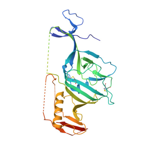 | |
UniProt | |||||
Find proteins for Q05320 (Zaire ebolavirus (strain Mayinga-76)) Explore Q05320 Go to UniProtKB: Q05320 | |||||
Entity Groups | |||||
| Sequence Clusters | 30% Identity50% Identity70% Identity90% Identity95% Identity100% Identity | ||||
| UniProt Group | Q05320 | ||||
Sequence AnnotationsExpand | |||||
| |||||
Find similar proteins by:
(by identity cutoff) | 3D Structure
Entity ID: 2 | |||||
|---|---|---|---|---|---|
| Molecule | Chains | Sequence Length | Organism | Details | Image |
| Envelope glycoprotein 2 | 168 | Ebola virus - Mayinga, Zaire, 1976 | Mutation(s): 0 Gene Names: GP |  | |
UniProt | |||||
Find proteins for Q05320 (Zaire ebolavirus (strain Mayinga-76)) Explore Q05320 Go to UniProtKB: Q05320 | |||||
Entity Groups | |||||
| Sequence Clusters | 30% Identity50% Identity70% Identity90% Identity95% Identity100% Identity | ||||
| UniProt Group | Q05320 | ||||
Sequence AnnotationsExpand | |||||
| |||||
Oligosaccharides
Entity ID: 3 | |||||
|---|---|---|---|---|---|
| Molecule | Chains | Length | 2D Diagram | Glycosylation | 3D Interactions |
| alpha-D-mannopyranose-(1-6)-beta-D-mannopyranose-(1-4)-2-acetamido-2-deoxy-beta-D-glucopyranose-(1-4)-2-acetamido-2-deoxy-beta-D-glucopyranose | C | 4 |  | N-Glycosylation | |
Glycosylation Resources | |||||
GlyTouCan: G22573RC GlyCosmos: G22573RC GlyGen: G22573RC | |||||
Small Molecules
| Ligands 4 Unique | |||||
|---|---|---|---|---|---|
| ID | Chains | Name / Formula / InChI Key | 2D Diagram | 3D Interactions | |
| T0R Query on T0R | K [auth B] | Toremifene C26 H28 Cl N O XFCLJVABOIYOMF-QPLCGJKRSA-N |  | ||
| NAG Query on NAG | D [auth A], E [auth A], F [auth A], G [auth A] | 2-acetamido-2-deoxy-beta-D-glucopyranose C8 H15 N O6 OVRNDRQMDRJTHS-FMDGEEDCSA-N |  | ||
| GOL Query on GOL | H [auth A], I [auth A] | GLYCEROL C3 H8 O3 PEDCQBHIVMGVHV-UHFFFAOYSA-N |  | ||
| DMS Query on DMS | J [auth A] | DIMETHYL SULFOXIDE C2 H6 O S IAZDPXIOMUYVGZ-UHFFFAOYSA-N |  | ||
Experimental Data & Validation
Experimental Data
- Method: X-RAY DIFFRACTION
- Resolution: 2.69 Å
- R-Value Free: 0.245
- R-Value Work: 0.203
- R-Value Observed: 0.205
- Space Group: H 3 2
Unit Cell:
| Length ( Å ) | Angle ( ˚ ) |
|---|---|
| a = 113.45 | α = 90 |
| b = 113.45 | β = 90 |
| c = 306.87 | γ = 120 |
| Software Name | Purpose |
|---|---|
| REFMAC | refinement |
| xia2 | data reduction |
| xia2 | data scaling |
| MOLREP | phasing |
Entry History
Deposition Data
- Released Date: 2016-06-29 Deposition Author(s): Zhao, Y., Ren, J., Stuart, D.I.
Revision History (Full details and data files)
- Version 1.0: 2016-06-29
Type: Initial release - Version 1.1: 2016-07-13
Changes: Database references - Version 2.0: 2020-07-29
Type: Remediation
Reason: Carbohydrate remediation
Changes: Atomic model, Data collection, Derived calculations, Structure summary - Version 2.1: 2024-01-10
Changes: Data collection, Database references, Derived calculations, Refinement description, Structure summary
