


Crystal structure of the serine endoprotease from Yersinia pestis
Filippova, E.V., Wawrzsak, Z., Sandoval, J., Skarina, T., Grimshaw, S., Savchenko, A., Anderson, W.F., Center for Structural Genomics of Infectious Diseases (CSGID)To be published.
Experimental Data Snapshot
Entity ID: 1 | |||||
|---|---|---|---|---|---|
| Molecule | Chains | Sequence Length | Organism | Details | Image |
| Periplasmic serine peptidase DegS | 334 | Yersinia pestis CO92 | Mutation(s): 0 Gene Names: degS, AK38_3169 | 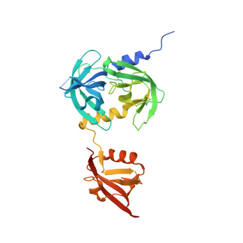 | |
UniProt | |||||
Find proteins for A0A5P8YL96 (Yersinia pestis) Explore A0A5P8YL96 Go to UniProtKB: A0A5P8YL96 | |||||
Entity Groups | |||||
| Sequence Clusters | 30% Identity50% Identity70% Identity90% Identity95% Identity100% Identity | ||||
| UniProt Group | A0A5P8YL96 | ||||
Sequence AnnotationsExpand | |||||
| |||||
| Ligands 3 Unique | |||||
|---|---|---|---|---|---|
| ID | Chains | Name / Formula / InChI Key | 2D Diagram | 3D Interactions | |
| CXS Query on CXS | B [auth A] | 3-CYCLOHEXYL-1-PROPYLSULFONIC ACID C9 H19 N O3 S PJWWRFATQTVXHA-UHFFFAOYSA-N |  | ||
| PEG Query on PEG | C [auth A] | DI(HYDROXYETHYL)ETHER C4 H10 O3 MTHSVFCYNBDYFN-UHFFFAOYSA-N |  | ||
| SO4 Query on SO4 | D [auth A] | SULFATE ION O4 S QAOWNCQODCNURD-UHFFFAOYSA-L |  | ||
| Length ( Å ) | Angle ( ˚ ) |
|---|---|
| a = 104.418 | α = 90 |
| b = 104.418 | β = 90 |
| c = 76.015 | γ = 120 |
| Software Name | Purpose |
|---|---|
| REFMAC | refinement |
| HKL-2000 | data scaling |
| PHENIX | phasing |
| Coot | model building |
RCSB PDB (citation) is hosted by
RCSB PDB is a member of the

