


STRUCTURE OF LIGAND BOUND CD33 RECEPTOR ASSOCIATED WITH ALZHEIMER'S DISEASE
Dodd, R.B., Meadows, W., Qamar, S., Johnson, C.M., Kronenberg-Versteeg, D., St George-Hyslop, P.To be published.
Experimental Data Snapshot
Entity ID: 1 | |||||
|---|---|---|---|---|---|
| Molecule | Chains | Sequence Length | Organism | Details | Image |
| Myeloid cell surface antigen CD33 | 224 | Homo sapiens | Mutation(s): 1 Gene Names: CD33, SIGLEC3 | 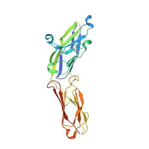 | |
UniProt & NIH Common Fund Data Resources | |||||
Find proteins for P20138 (Homo sapiens) Explore P20138 Go to UniProtKB: P20138 | |||||
PHAROS: P20138 GTEx: ENSG00000105383 | |||||
Entity Groups | |||||
| Sequence Clusters | 30% Identity50% Identity70% Identity90% Identity95% Identity100% Identity | ||||
| UniProt Group | P20138 | ||||
Sequence AnnotationsExpand | |||||
| |||||
| Ligands 2 Unique | |||||
|---|---|---|---|---|---|
| ID | Chains | Name / Formula / InChI Key | 2D Diagram | 3D Interactions | |
| NAG Query on NAG | H [auth A] I [auth A] J [auth A] K [auth B] L [auth B] | 2-acetamido-2-deoxy-beta-D-glucopyranose C8 H15 N O6 OVRNDRQMDRJTHS-FMDGEEDCSA-N |  | ||
| PG0 Query on PG0 | Q [auth C], R [auth C] | 2-(2-METHOXYETHOXY)ETHANOL C5 H12 O3 SBASXUCJHJRPEV-UHFFFAOYSA-N |  | ||
| Length ( Å ) | Angle ( ˚ ) |
|---|---|
| a = 64.49 | α = 90 |
| b = 123.68 | β = 90 |
| c = 141.6 | γ = 90 |
| Software Name | Purpose |
|---|---|
| PHENIX | refinement |
| xia2 | data reduction |
| XSCALE | data scaling |
| XDS | data reduction |
| PHASER | phasing |
| Funding Organization | Location | Grant Number |
|---|---|---|
| Wellcome Trust | United Kingdom | RG47376 |
RCSB PDB (citation) is hosted by
RCSB PDB is a member of the


