5IKN

Crystal Structure of the T7 Replisome in the Absence of DNA


Experimental Data Snapshot

  • Method: X-RAY DIFFRACTION
  • Resolution: 4.80 Å
  • R-Value Free: 0.318 
  • R-Value Work: 0.275 
  • R-Value Observed: 0.277 

wwPDB Validation   3D Report Full Report


This is version 1.4 of the entry. See complete history


Literature

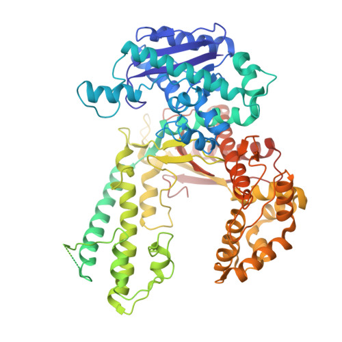
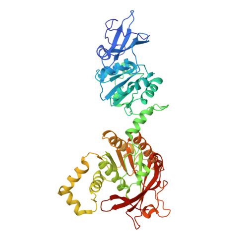
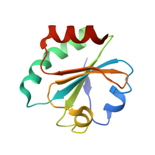
Hybrid Methods Reveal Multiple Flexibly Linked DNA Polymerases within the Bacteriophage T7 Replisome.

Wallen, J.R.Zhang, H.Weis, C.Cui, W.Foster, B.M.Ho, C.M.Hammel, M.Tainer, J.A.Gross, M.L.Ellenberger, T.

(2017) Structure 25: 157-166

  • DOI: https://doi.org/10.1016/j.str.2016.11.019
  • Primary Citation of Related Structures:  
    5IKN

  • PubMed Abstract: 

    The physical organization of DNA enzymes at a replication fork enables efficient copying of two antiparallel DNA strands, yet dynamic protein interactions within the replication complex complicate replisome structural studies. We employed a combination of crystallographic, native mass spectrometry and small-angle X-ray scattering experiments to capture alternative structures of a model replication system encoded by bacteriophage T7. Two molecules of DNA polymerase bind the ring-shaped primase-helicase in a conserved orientation and provide structural insight into how the acidic C-terminal tail of the primase-helicase contacts the DNA polymerase to facilitate loading of the polymerase onto DNA. A third DNA polymerase binds the ring in an offset manner that may enable polymerase exchange during replication. Alternative polymerase binding modes are also detected by small-angle X-ray scattering with DNA substrates present. Our collective results unveil complex motions within T7 replisome higher-order structures that are underpinned by multivalent protein-protein interactions with functional implications.


  • Organizational Affiliation

    Department of Chemistry & Physics, Western Carolina University, Cullowhee, NC 28723, USA. Electronic address: jamiewallen@email.wcu.edu.


Macromolecules
Find similar proteins by:  (by identity cutoff)  |  3D Structure
Entity ID: 1
MoleculeChains Sequence LengthOrganismDetailsImage
DNA-directed DNA polymerase
A, B, C
704Escherichia phage T7Mutation(s): 2 
EC: 2.7.7.7 (PDB Primary Data), 3.1.11 (PDB Primary Data)
UniProt
Find proteins for P00581 (Escherichia phage T7)
Explore P00581 
Go to UniProtKB:  P00581
Entity Groups  
Sequence Clusters30% Identity50% Identity70% Identity90% Identity95% Identity100% Identity
UniProt GroupP00581
Sequence Annotations
Expand
  • Reference Sequence
Find similar proteins by:  (by identity cutoff)  |  3D Structure
Entity ID: 2
MoleculeChains Sequence LengthOrganismDetailsImage
DNA primase/helicase
D, E, F, G, H
D, E, F, G, H, I, J
486Escherichia phage T7Mutation(s): 0 
EC: 2.7.7 (PDB Primary Data), 3.6.4.12 (PDB Primary Data)
UniProt
Find proteins for P03692 (Escherichia phage T7)
Explore P03692 
Go to UniProtKB:  P03692
Entity Groups  
Sequence Clusters30% Identity50% Identity70% Identity90% Identity95% Identity100% Identity
UniProt GroupP03692
Sequence Annotations
Expand
  • Reference Sequence
Find similar proteins by:  (by identity cutoff)  |  3D Structure
Entity ID: 3
MoleculeChains Sequence LengthOrganismDetailsImage
Thioredoxin-1
K, L, M
105Escherichia coli O157:H7Mutation(s): 0 
Gene Names: trxAZ5291ECs4714
UniProt
Find proteins for P0AA27 (Escherichia coli O157:H7)
Explore P0AA27 
Go to UniProtKB:  P0AA27
Entity Groups  
Sequence Clusters30% Identity50% Identity70% Identity90% Identity95% Identity100% Identity
UniProt GroupP0AA27
Sequence Annotations
Expand
  • Reference Sequence
Experimental Data & Validation

Experimental Data

  • Method: X-RAY DIFFRACTION
  • Resolution: 4.80 Å
  • R-Value Free: 0.318 
  • R-Value Work: 0.275 
  • R-Value Observed: 0.277 
  • Space Group: P 21 21 21
Unit Cell:
Length ( Å )Angle ( ˚ )
a = 174.866α = 90
b = 238.091β = 90
c = 243.466γ = 90
Software Package:
Software NamePurpose
PHENIXrefinement
HKL-2000data scaling
PDB_EXTRACTdata extraction
HKL-2000data reduction

Structure Validation

View Full Validation Report


Entry History & Funding Information

Deposition Data


Funding OrganizationLocationGrant Number
National Institutes of Health/National Institute of General Medical Sciences (NIH/NIGMS)United StatesGM055390

Revision History  (Full details and data files)

  • Version 1.0: 2016-12-07
    Type: Initial release
  • Version 1.1: 2017-01-18
    Changes: Database references
  • Version 1.2: 2017-09-20
    Changes: Author supporting evidence
  • Version 1.3: 2019-12-25
    Changes: Author supporting evidence
  • Version 1.4: 2023-09-27
    Changes: Data collection, Database references, Refinement description