5I8L

Crystal structure of the full-length cell wall-binding module of Cpl7 mutant R223A


Experimental Data Snapshot

  • Method: X-RAY DIFFRACTION
  • Resolution: 2.80 Å
  • R-Value Free: 0.289 
  • R-Value Work: 0.206 
  • R-Value Observed: 0.209 

wwPDB Validation   3D Report Full Report


This is version 1.2 of the entry. See complete history


Literature

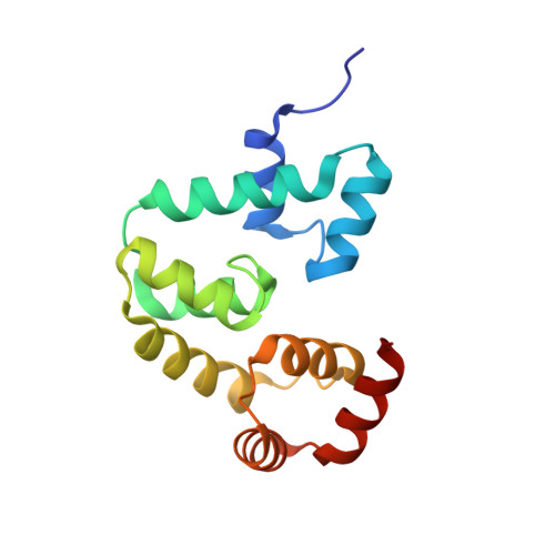
Deciphering how Cpl-7 cell wall-binding repeats recognize the bacterial peptidoglycan.

Bustamante, N.Iglesias-Bexiga, M.Bernardo-Garcia, N.Silva-Martin, N.Garcia, G.Campanero-Rhodes, M.A.Garcia, E.Uson, I.Buey, R.M.Garcia, P.Hermoso, J.A.Bruix, M.Menendez, M.

(2017) Sci Rep 7: 16494-16494

  • DOI: https://doi.org/10.1038/s41598-017-16392-4
  • Primary Citation of Related Structures:  
    4CVD, 5I8L

  • PubMed Abstract: 

    Endolysins, the cell wall lytic enzymes encoded by bacteriophages to release the phage progeny, are among the top alternatives to fight against multiresistant pathogenic bacteria; one of the current biggest challenges to global health. Their narrow range of susceptible bacteria relies, primarily, on targeting specific cell-wall receptors through specialized modules. The cell wall-binding domain of Cpl-7 endolysin, made of three CW_7 repeats, accounts for its extended-range of substrates. Using as model system the cell wall-binding domain of Cpl-7, here we describe the molecular basis for the bacterial cell wall recognition by the CW_7 motif, which is widely represented in sequences of cell wall hydrolases. We report the crystal and solution structure of the full-length domain, identify N-acetyl-D-glucosaminyl-(β1,4)-N-acetylmuramyl-L-alanyl-D-isoglutamine (GMDP) as the peptidoglycan (PG) target recognized by the CW_7 motifs, and characterize feasible GMDP-CW_7 contacts. Our data suggest that Cpl-7 cell wall-binding domain might simultaneously bind to three PG chains, and also highlight the potential use of CW_7-containing lysins as novel anti-infectives.


  • Organizational Affiliation

    Instituto de Química-Física Rocasolano, Consejo Superior de Investigaciones Científicas, Serrano 119, 28006, Madrid, Spain.


Macromolecules
Find similar proteins by:  (by identity cutoff)  |  3D Structure
Entity ID: 1
MoleculeChains Sequence LengthOrganismDetailsImage
Lysozyme145Streptococcus phage CP-7Mutation(s): 3 
Gene Names: CPL7
EC: 3.2.1.17
UniProt
Find proteins for P19385 (Streptococcus phage Cp-7)
Explore P19385 
Go to UniProtKB:  P19385
Entity Groups  
Sequence Clusters30% Identity50% Identity70% Identity90% Identity95% Identity100% Identity
UniProt GroupP19385
Sequence Annotations
Expand
  • Reference Sequence
Small Molecules
Ligands 1 Unique
IDChains Name / Formula / InChI Key2D Diagram3D Interactions
GOL
Query on GOL

Download Ideal Coordinates CCD File 
B [auth A]GLYCEROL
C3 H8 O3
PEDCQBHIVMGVHV-UHFFFAOYSA-N
Experimental Data & Validation

Experimental Data

  • Method: X-RAY DIFFRACTION
  • Resolution: 2.80 Å
  • R-Value Free: 0.289 
  • R-Value Work: 0.206 
  • R-Value Observed: 0.209 
  • Space Group: P 21 21 21
Unit Cell:
Length ( Å )Angle ( ˚ )
a = 29.524α = 90
b = 50.331β = 90
c = 86.089γ = 90
Software Package:
Software NamePurpose
PHENIXrefinement
XDSdata reduction
Aimlessdata scaling
Cootmodel building

Structure Validation

View Full Validation Report


Entry History 

Deposition Data

Revision History  (Full details and data files)

  • Version 1.0: 2017-03-08
    Type: Initial release
  • Version 1.1: 2017-12-20
    Changes: Database references
  • Version 1.2: 2024-01-10
    Changes: Data collection, Database references, Refinement description