Structural characterizations of phage antitoxin Dmd and its interactions with bacterial toxin RnlA
Wei, Y., Gao, Z.Q., Zhang, H., Dong, Y.H.(2016) Biochem Biophys Res Commun 472: 592-597
- PubMed: 26972252
- DOI: https://doi.org/10.1016/j.bbrc.2016.03.025
- Primary Citation of Related Structures:
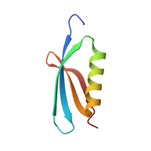
5I8J - PubMed Abstract:
Toxin-antitoxin (TA) loci are widespread in bacteria plasmids and chromosomes, and target various cellular functions to regulate cell growth and death. A type II TA system RnlA-RnlB from Escherichia coli is associated with phage-resistance. After the infection of bacteriophage T4 with Dmd defection, RnlA is activated by the disappearance of RnlB, resulting in the rapid degradation of T4 mRNAs. Dmd can bind to RnlA directly and neutralize RnlA toxicity to allow phage reproduction. Dmd represent a heterogenous antitoxin of RnlA replacing antitoxin RnlB. Here, we reported two structures of Dmd from T4 phage and RB69 phage. Both Dmd structures are high similar with a compacted domain composed of a four-stranded anti-parallel β-sheet and an α-helix. Chromatography and SAXS suggest Dmd forms a dimer in solution consistent with that in crystal. Structure-based mutagenesis of Dmd reveals key residues involved in RnlA-binding. Possibility cavities in Dmd used for compounds design were modeled. Our structural study revealed the recognition and inhibition mechanism of RnlA by Dmd and providing a potential laboratory phage prevention target for drug design.
Organizational Affiliation:
Beijing Synchrotron Radiation Facility, Institute of High Energy Physics, Chinese Academy of Sciences, Beijing, 100049, China.