5HY3

Crystal structure of Escherichia coli toxin LsoA in complex with T4 phage antitoxin Dmd


Experimental Data Snapshot

  • Method: X-RAY DIFFRACTION
  • Resolution: 3.10 Å
  • R-Value Free: 0.302 
  • R-Value Work: 0.259 
  • R-Value Observed: 0.262 

wwPDB Validation   3D Report Full Report


This is version 1.2 of the entry. See complete history


Literature

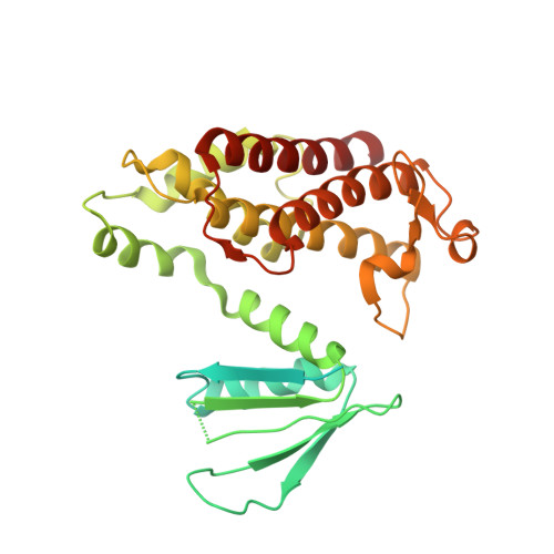
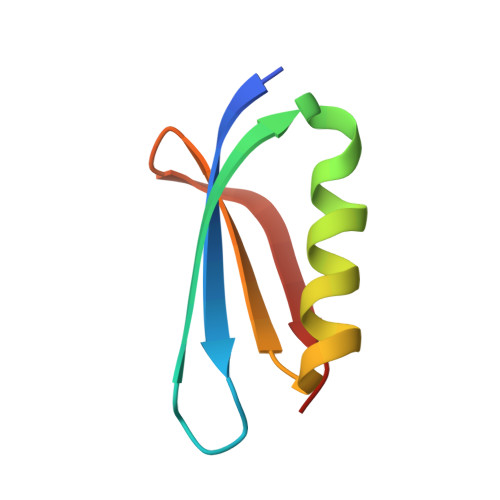
Structural insights into the inhibition mechanism of bacterial toxin LsoA by bacteriophage antitoxin Dmd

Wan, H.Otsuka, Y.Gao, Z.Q.Wei, Y.Chen, Z.Masuda, M.Yonesaki, T.Zhang, H.Dong, Y.H.

(2016) Mol Microbiol 101: 757-769

  • DOI: https://doi.org/10.1111/mmi.13420
  • Primary Citation of Related Structures:  
    5HY3

  • PubMed Abstract: 

    Bacteria have obtained a variety of resistance mechanisms including toxin-antitoxin (TA) systems against bacteriophages (phages), whereas phages have also evolved to overcome bacterial anti-phage mechanisms. Dmd from T4 phage can suppress the toxicities of homologous toxins LsoA and RnlA from Escherichia coli, representing the first example of a phage antitoxin against multiple bacterial toxins in known TA systems. Here, the crystal structure of LsoA-Dmd complex showed Dmd is inserted into the deep groove between the N-terminal repeated domain (NRD) and the Dmd-binding domain (DBD) of LsoA. The NRD shifts significantly from a 'closed' to an 'open' conformation upon Dmd binding. Site-directed mutagenesis of Dmd revealed the conserved residues (W31 and N40) are necessary for LsoA binding and the toxicity suppression as determined by pull-down and cell toxicity assays. Further mutagenesis identified the conserved Dmd-binding residues (R243, E246 and R305) of LsoA are vital for its toxicity, and suggested Dmd and LsoB may possess different inhibitory mechanisms against LsoA toxicity. Our structure-function studies demonstrate Dmd can recognize LsoA and inhibit its toxicity by occupying the active site possibly via substrate mimicry. These findings have provided unique insights into the defense and counter-defense mechanisms between bacteria and phages in their co-evolution.


  • Organizational Affiliation

    Beijing Synchrotron Radiation Facility, Institute of High Energy Physics, Chinese Academy of Sciences, Beijing, 100049, China.


Macromolecules
Find similar proteins by:  (by identity cutoff)  |  3D Structure
Entity ID: 1
MoleculeChains Sequence LengthOrganismDetailsImage
mRNA endoribonuclease LsoA346Escherichia coli O157:H7Mutation(s): 0 
Gene Names: lsoA
EC: 3.1
UniProt
Find proteins for O82881 (Escherichia coli O157:H7)
Explore O82881 
Go to UniProtKB:  O82881
Entity Groups  
Sequence Clusters30% Identity50% Identity70% Identity90% Identity95% Identity100% Identity
UniProt GroupO82881
Sequence Annotations
Expand
  • Reference Sequence
Find similar proteins by:  (by identity cutoff)  |  3D Structure
Entity ID: 2
MoleculeChains Sequence LengthOrganismDetailsImage
Antitoxin Dmd60Tequatrovirus T4Mutation(s): 0 
Gene Names: dmd61.5y02B
UniProt
Find proteins for P39232 (Enterobacteria phage T4)
Explore P39232 
Go to UniProtKB:  P39232
Entity Groups  
Sequence Clusters30% Identity50% Identity70% Identity90% Identity95% Identity100% Identity
UniProt GroupP39232
Sequence Annotations
Expand
  • Reference Sequence
Experimental Data & Validation

Experimental Data

  • Method: X-RAY DIFFRACTION
  • Resolution: 3.10 Å
  • R-Value Free: 0.302 
  • R-Value Work: 0.259 
  • R-Value Observed: 0.262 
  • Space Group: P 41 21 2
Unit Cell:
Length ( Å )Angle ( ˚ )
a = 112.431α = 90
b = 112.431β = 90
c = 95.083γ = 90
Software Package:
Software NamePurpose
PHENIXrefinement
HKL-2000data reduction
HKL-2000data scaling
PHASERphasing

Structure Validation

View Full Validation Report


Entry History 

Deposition Data

Revision History  (Full details and data files)

  • Version 1.0: 2016-06-22
    Type: Initial release
  • Version 1.1: 2016-09-07
    Changes: Database references
  • Version 1.2: 2024-04-03
    Changes: Data collection, Database references, Derived calculations, Refinement description