5HWN

Crystal structure of keto-deoxy-D-galactarate dehydratase complexed with pyruvate


Experimental Data Snapshot

  • Method: X-RAY DIFFRACTION
  • Resolution: 1.50 Å
  • R-Value Free: 0.170 
  • R-Value Work: 0.144 
  • R-Value Observed: 0.146 

wwPDB Validation   3D Report Full Report


This is version 2.0 of the entry. See complete history

Re-refinement Note

This entry reflects an alternative modeling of the original data in: 4ur7


Literature

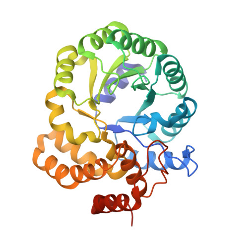
Structure and function of a decarboxylating Agrobacterium tumefaciens keto-deoxy-d-galactarate dehydratase.

Taberman, H.Andberg, M.Parkkinen, T.Janis, J.Penttila, M.Hakulinen, N.Koivula, A.Rouvinen, J.

(2014) Biochemistry 53: 8052-8060

  • DOI: https://doi.org/10.1021/bi501290k
  • Primary Citation of Related Structures:  
    4UR7, 4UR8, 5HWJ, 5HWM, 5HWN

  • PubMed Abstract: 

    Agrobacterium tumefaciens (At) strain C58 contains an oxidative enzyme pathway that can function on both d-glucuronic and d-galacturonic acid. The corresponding gene coding for At keto-deoxy-d-galactarate (KDG) dehydratase is located in the same gene cluster as those coding for uronate dehydrogenase (At Udh) and galactarolactone cycloisomerase (At Gci) which we have previously characterized. Here, we present the kinetic characterization and crystal structure of At KDG dehydratase, which catalyzes the next step, the decarboxylating hydrolyase reaction of KDG to produce α-ketoglutaric semialdehyde (α-KGSA) and carbon dioxide. The crystal structures of At KDG dehydratase and its complexes with pyruvate and 2-oxoadipic acid, two substrate analogues, were determined to 1.7 Å, 1.5 Å, and 2.1 Å resolution, respectively. Furthermore, mass spectrometry was used to confirm reaction end-products. The results lead us to propose a structure-based mechanism for At KDG dehydratase, suggesting that while the enzyme belongs to the Class I aldolase protein family, it does not follow a typical retro-aldol condensation mechanism.


  • Organizational Affiliation

    Department of Chemistry, University of Eastern Finland , FI-80101 Joensuu, Finland.


Macromolecules
Find similar proteins by:  (by identity cutoff)  |  3D Structure
Entity ID: 1
MoleculeChains Sequence LengthOrganismDetailsImage
Probable 5-dehydro-4-deoxyglucarate dehydratase
A, B, C, D
311Agrobacterium fabrum str. C58Mutation(s): 0 
Gene Names: Atu3140AGR_L_3338
EC: 4.2.1.41
UniProt
Find proteins for Q8UB77 (Agrobacterium fabrum (strain C58 / ATCC 33970))
Explore Q8UB77 
Go to UniProtKB:  Q8UB77
Entity Groups  
Sequence Clusters30% Identity50% Identity70% Identity90% Identity95% Identity100% Identity
UniProt GroupQ8UB77
Sequence Annotations
Expand
  • Reference Sequence
Small Molecules
Ligands 3 Unique
IDChains Name / Formula / InChI Key2D Diagram3D Interactions
GOL
Query on GOL

Download Ideal Coordinates CCD File 
F [auth A]
G [auth A]
H [auth A]
I [auth A]
K [auth A]
F [auth A],
G [auth A],
H [auth A],
I [auth A],
K [auth A],
M [auth B],
N [auth B],
O [auth B],
R [auth C],
U [auth D],
V [auth D],
W [auth D]
GLYCEROL
C3 H8 O3
PEDCQBHIVMGVHV-UHFFFAOYSA-N
PYR
Query on PYR

Download Ideal Coordinates CCD File 
E [auth A],
L [auth B],
Q [auth C],
T [auth D]
PYRUVIC ACID
C3 H4 O3
LCTONWCANYUPML-UHFFFAOYSA-N
FMT
Query on FMT

Download Ideal Coordinates CCD File 
J [auth A],
P [auth B],
S [auth C],
X [auth D]
FORMIC ACID
C H2 O2
BDAGIHXWWSANSR-UHFFFAOYSA-N
Experimental Data & Validation

Experimental Data

  • Method: X-RAY DIFFRACTION
  • Resolution: 1.50 Å
  • R-Value Free: 0.170 
  • R-Value Work: 0.144 
  • R-Value Observed: 0.146 
  • Space Group: C 1 2 1
Unit Cell:
Length ( Å )Angle ( ˚ )
a = 169.8α = 90
b = 120.14β = 112.01
c = 74.07γ = 90
Software Package:
Software NamePurpose
PHENIXrefinement
XDSdata reduction
XSCALEdata scaling
PHASERphasing

Structure Validation

View Full Validation Report


Entry History 

Deposition Data

Revision History  (Full details and data files)

  • Version 1.0: 2016-03-23
    Type: Initial release
  • Version 2.0: 2023-11-15
    Changes: Atomic model, Data collection, Database references, Derived calculations