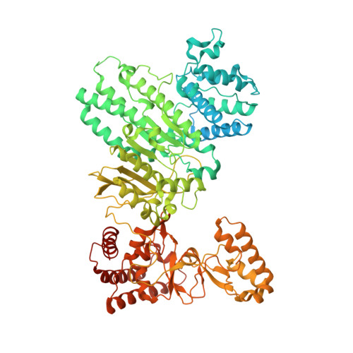
5HR4

Structure of Type IIL restriction-modification enzyme MmeI in complex with DNA has implications for engineering of new specificities


Experimental Data Snapshot

  • Method: X-RAY DIFFRACTION
  • Resolution: 2.60 Å
  • R-Value Free: 0.242 
  • R-Value Work: 0.215 
  • R-Value Observed: 0.216 

wwPDB Validation   3D Report Full Report


Ligand Structure Quality Assessment 


This is version 1.1 of the entry. See complete history


Literature

Structure of Type IIL Restriction-Modification Enzyme MmeI in Complex with DNA Has Implications for Engineering New Specificities.

Callahan, S.J.Luyten, Y.A.Gupta, Y.K.Wilson, G.G.Roberts, R.J.Morgan, R.D.Aggarwal, A.K.

(2016) PLoS Biol 14: e1002442-e1002442

  • DOI: https://doi.org/10.1371/journal.pbio.1002442
  • Primary Citation of Related Structures:  
    5HR4

  • PubMed Abstract: 

    The creation of restriction enzymes with programmable DNA-binding and -cleavage specificities has long been a goal of modern biology. The recently discovered Type IIL MmeI family of restriction-and-modification (RM) enzymes that possess a shared target recognition domain provides a framework for engineering such new specificities. However, a lack of structural information on Type IIL enzymes has limited the repertoire that can be rationally engineered. We report here a crystal structure of MmeI in complex with its DNA substrate and an S-adenosylmethionine analog (Sinefungin). The structure uncovers for the first time the interactions that underlie MmeI-DNA recognition and methylation (5'-TCCRAC-3'; R = purine) and provides a molecular basis for changing specificity at four of the six base pairs of the recognition sequence (5'-TCCRAC-3'). Surprisingly, the enzyme is resilient to specificity changes at the first position of the recognition sequence (5'-TCCRAC-3'). Collectively, the structure provides a basis for engineering further derivatives of MmeI and delineates which base pairs of the recognition sequence are more amenable to alterations than others.


  • Organizational Affiliation

    Department of Structural and Chemical Biology, Mount Sinai School of Medicine, New York, New York, United States of America.


Macromolecules

Find similar proteins by:  (by identity cutoff)  |  3D Structure
Entity ID: 1
MoleculeChains Sequence LengthOrganismDetailsImage
MmeIA [auth C],
D [auth J]
919Methylophilus methylotrophusMutation(s): 0 
Gene Names: mmeIRM
UniProt
Find proteins for B2MU09 (Methylophilus methylotrophus)
Explore B2MU09 
Go to UniProtKB:  B2MU09
Entity Groups  
Sequence Clusters30% Identity50% Identity70% Identity90% Identity95% Identity100% Identity
UniProt GroupB2MU09
Sequence Annotations
Expand
  • Reference Sequence

Find similar nucleic acids by:  Sequence   |   3D Structure  

Entity ID: 2
MoleculeChains LengthOrganismImage
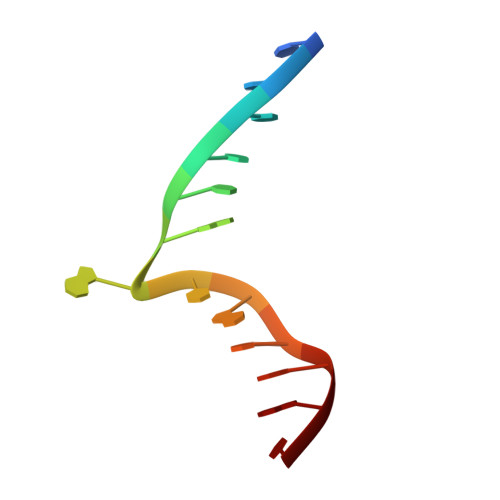
DNA (5'-D(P*TP*AP*TP*CP*CP*GP*AP*CP*AP*TP*AP*AP*C)-3')B [auth H],
E [auth K]
13Methylophilus methylotrophus
Sequence Annotations
Expand
  • Reference Sequence

Find similar nucleic acids by:  Sequence   |   3D Structure  

Entity ID: 3
MoleculeChains LengthOrganismImage
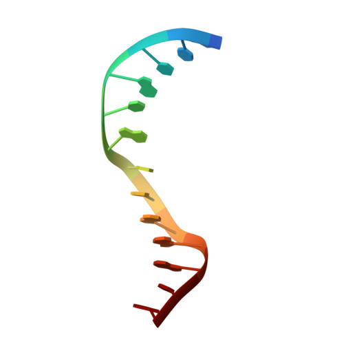
DNA (5'-D(P*GP*TP*TP*AP*TP*GP*TP*CP*GP*GP*AP*TP*A)-3')C [auth I],
F [auth L]
13Methylophilus methylotrophus
Sequence Annotations
Expand
  • Reference Sequence
Experimental Data & Validation

Experimental Data

  • Method: X-RAY DIFFRACTION
  • Resolution: 2.60 Å
  • R-Value Free: 0.242 
  • R-Value Work: 0.215 
  • R-Value Observed: 0.216 
  • Space Group: P 1
Unit Cell:
Length ( Å )Angle ( ˚ )
a = 61.877α = 72.84
b = 95.292β = 89.15
c = 161.969γ = 71.62
Software Package:
Software NamePurpose
PHENIXrefinement
XDSdata reduction
XDSdata scaling
PHENIXphasing

Structure Validation

View Full Validation Report


Ligand Structure Quality Assessment 


Entry History 

Deposition Data

Revision History  (Full details and data files)

  • Version 1.0: 2016-04-27
    Type: Initial release
  • Version 1.1: 2024-03-06
    Changes: Data collection, Database references, Derived calculations