Monomeric Form of Peptidylarginine Deiminase Type I Revealed by X-ray Crystallography and Small-Angle X-ray Scattering
Saijo, S., Nagai, A., Kinjo, S., Mashimo, R., Akimoto, M., Kizawa, K., Yabe-Wada, T., Shimizu, N., Takahara, H., Unno, M.(2016) J Mol Biol 428: 3058-3073
- PubMed: 27393304
- DOI: https://doi.org/10.1016/j.jmb.2016.06.018
- Primary Citation of Related Structures:
5HP5 - PubMed Abstract:
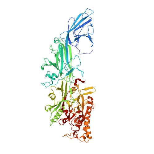
Peptidylarginine deiminase (PAD; EC 3.5.3.15) is a post-translational modification enzyme that catalyzes the conversion of arginine in protein molecules to a citrulline residue in a Ca(2+)-dependent manner. In this study, we determined the structure of an active form of human PAD1 crystallized in the presence of Ca(2+) at 3.2-Å resolution. Although human PAD2 and PAD4 isozymes were previously reported to form a head-to-tail homodimer, it is still unknown whether this quaternary structure is common to other PAD isozymes. The asymmetric unit of the crystal contained two PAD1 molecules; however, the head-to-tail dimeric form was not found. Small-angle X-ray scattering analyses revealed PAD1 to be a monomer in solution, while PAD3 was dimerized with a structure similar to PAD2 and PAD4. PAD1 was apparently different from the crystal structures of PAD2 and PAD4, with an elongated N-terminal loop that appears to prevent the formation of the homodimer. Of interest, the N-terminal loop occupied the substrate binding site of the adjacent PAD1 molecules in the crystal. Deimination of S100A3 peptides in vitro implied that PAD isozymes recognize the quaternary structure of S100A3. The substrate-accessible monomeric structure brought about by the extension of its N terminus may partly account for the highest tolerant substrate recognition of PAD1. This is the first ever report on the molecular structure of PAD1 demonstrating the unique monomeric form of the PAD isozyme.
Organizational Affiliation:
Institute of Materials Structure Science, High Energy Accelerator Research Organization, KEK, 1-1 Oho, Tsukuba, Ibaraki 305-0801, Japan.