5HNP

The structure of the kdo-capped saccharide binding subunit of the O-12 specific ABC transporter, Wzt


Experimental Data Snapshot

  • Method: X-RAY DIFFRACTION
  • Resolution: 2.20 Å
  • R-Value Free: 0.219 
  • R-Value Work: 0.193 
  • R-Value Observed: 0.194 

wwPDB Validation   3D Report Full Report


This is version 1.4 of the entry. See complete history


Literature

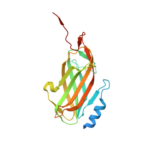
The Klebsiella pneumoniae O12 ATP-binding Cassette (ABC) Transporter Recognizes the Terminal Residue of Its O-antigen Polysaccharide Substrate.

Mann, E.Mallette, E.Clarke, B.R.Kimber, M.S.Whitfield, C.

(2016) J Biol Chem 291: 9748-9761

  • DOI: https://doi.org/10.1074/jbc.M116.719344
  • Primary Citation of Related Structures:  
    5HNO, 5HNP

  • PubMed Abstract: 

    Export of the Escherichia coli serotype O9a O-antigenic polysaccharides (O-PS) involves an ATP-binding cassette (ABC) transporter. The process requires a non-reducing terminal residue, which is recognized by a carbohydrate-binding module (CBM) appended to the C terminus of the nucleotide-binding domain of the transporter. Here, we investigate the process in Klebsiella pneumoniae serotype O12 (and Raoultella terrigena ATCC 33257). The O12 polysaccharide is terminated at the non-reducing end by a β-linked 3-deoxy-d-manno-oct-2-ulosonic acid (Kdo) residue. The O12 ABC transporter also binds its cognate O-PS via a CBM, and export is dependent on the presence of the terminal β-Kdo residue. The overall structural architecture of the O12 CBM resembles the O9a prototype, but they share only weak sequence similarity, and the putative binding pocket for the O12 glycan is different. Removal of the CBM abrogated O-PS transport, but export was restored when the CBM was expressed in trans with the mutant CBM-deficient ABC transporter. These results demonstrate that the CBM-mediated substrate-recognition mechanism is evolutionarily conserved and can operate with glycans of widely differing structures.


  • Organizational Affiliation

    From the Department of Molecular and Cellular Biology, University of Guelph, Guelph, Ontario N1G 2W1, Canada.


Macromolecules
Find similar proteins by:  (by identity cutoff)  |  3D Structure
Entity ID: 1
MoleculeChains Sequence LengthOrganismDetailsImage
ABC transporter
A, B
190Raoultella ornithinolyticaMutation(s): 0 
Gene Names: TE10_19180
Entity Groups  
Sequence Clusters30% Identity50% Identity70% Identity90% Identity95% Identity100% Identity
Sequence Annotations
Expand
  • Reference Sequence
Small Molecules
Ligands 1 Unique
IDChains Name / Formula / InChI Key2D Diagram3D Interactions
CL
Query on CL

Download Ideal Coordinates CCD File 
C [auth A]CHLORIDE ION
Cl
VEXZGXHMUGYJMC-UHFFFAOYSA-M
Experimental Data & Validation

Experimental Data

  • Method: X-RAY DIFFRACTION
  • Resolution: 2.20 Å
  • R-Value Free: 0.219 
  • R-Value Work: 0.193 
  • R-Value Observed: 0.194 
  • Space Group: P 21 21 21
Unit Cell:
Length ( Å )Angle ( ˚ )
a = 47.78α = 90
b = 104.07β = 90
c = 106.33γ = 90
Software Package:
Software NamePurpose
PHENIXrefinement
XDSdata reduction
XSCALEdata scaling
PHASERphasing

Structure Validation

View Full Validation Report


Entry History 

Deposition Data

Revision History  (Full details and data files)

  • Version 1.0: 2016-03-09
    Type: Initial release
  • Version 1.1: 2016-03-16
    Changes: Database references
  • Version 1.2: 2016-05-11
    Changes: Database references
  • Version 1.3: 2016-07-06
    Changes: Other
  • Version 1.4: 2023-09-27
    Changes: Data collection, Database references, Derived calculations, Refinement description