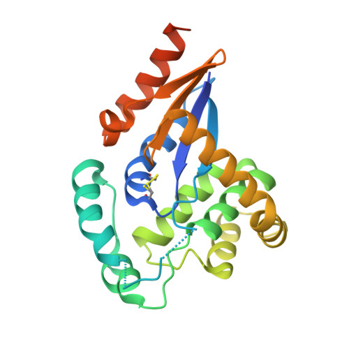
Crystal structures of the disulfide reductase DsbM from Pseudomonas aeruginosa
Jo, I., Park, N., Chung, I.Y., Cho, Y.H., Ha, N.-C.(2016) Acta Crystallogr D Struct Biol 72: 1100-1109
- PubMed: 27710931
- DOI: https://doi.org/10.1107/S2059798316013024
- Primary Citation of Related Structures:
5HFG, 5HFI - PubMed Abstract:
In bacteria, many Dsb-family proteins play diverse roles in the conversion between the oxidized and reduced states of cysteine residues of substrate proteins. Most Dsb enzymes catalyze disulfide formation in periplasmic or secreted substrate proteins. Recently, a DsbM protein has been found in a Gram-negative bacterium, and was characterized as a cytosolic Dsb member with the conserved CXXC motif on the basis of sequence homology to the Dsb-family proteins. The protein was implicated in the reduction of the cytoplasmic redox-sensor protein OxyR in Pseudomonas aeruginosa. Here, crystal structures of DsbM from P. aeruginosa are presented, revealing that it consists of a modified thioredoxin domain containing the CXXC motif and a lid domain surrounding the CXXC motif. In a glutathione-linked structure, a glutathione molecule is linked to the CXXC motif of DsbM and is bound in an elongated cavity region in the thioredoxin domain, which is also suited for substrate peptide binding. A striking structural similarity to a human glutathione S-transferase was found in the glutathione-binding pocket. Further, biochemical evidence is presented suggesting that DsbM is directly involved in the reduction of the disulfide of Cys199 and Cys208 in OxyR, resulting in the acceleration of OxyR reduction in the absence of reactive oxygen species stress. These findings may help to expand the understanding of the diverse roles of redox-related proteins that contain the CXXC motif.
Organizational Affiliation:
Department of Agricultural Biotechnology, Center for Food Safety and Toxicology, Center for Food and Bioconvergence, Research Institute for Agriculture and Life Sciences, Seoul National University, Seoul 08826, Republic of Korea.