5G1M

Crystal structure of NagZ from Pseudomonas aeruginosa


Experimental Data Snapshot

  • Method: X-RAY DIFFRACTION
  • Resolution: 1.80 Å
  • R-Value Free: 0.241 
  • R-Value Work: 0.196 
  • R-Value Observed: 0.198 

wwPDB Validation   3D Report Full Report


This is version 1.4 of the entry. See complete history


Literature

Catalytic Cycle of the N-Acetylglucosaminidase NagZ from Pseudomonas aeruginosa.

Acebron, I.Mahasenan, K.V.De Benedetti, S.Lee, M.Artola-Recolons, C.Hesek, D.Wang, H.Hermoso, J.A.Mobashery, S.

(2017) J Am Chem Soc 139: 6795-6798

  • DOI: https://doi.org/10.1021/jacs.7b01626
  • Primary Citation of Related Structures:  
    5G1M, 5G5K, 5G5U, 5G6T, 5LY7

  • PubMed Abstract: 

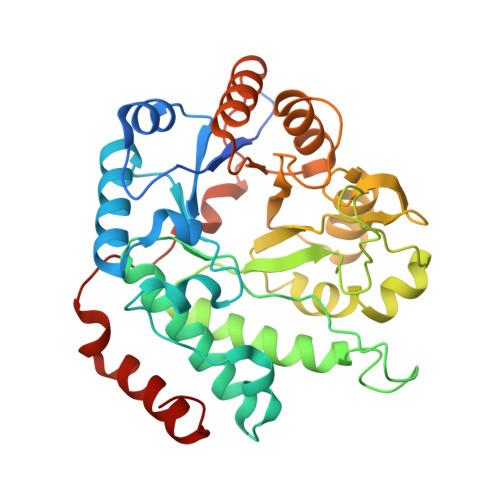
    The N-acetylglucosaminidase NagZ of Pseudomonas aeruginosa catalyzes the first cytoplasmic step in recycling of muropeptides, cell-wall-derived natural products. This reaction regulates gene expression for the β-lactam resistance enzyme, β-lactamase. The enzyme catalyzes hydrolysis of N-acetyl-β-d-glucosamine-(1→4)-1,6-anhydro-N-acetyl-β-d-muramyl-peptide (1) to N-acetyl-β-d-glucosamine (2) and 1,6-anhydro-N-acetyl-β-d-muramyl-peptide (3). The structural and functional aspects of catalysis by NagZ were investigated by a total of seven X-ray structures, three computational models based on the X-ray structures, molecular-dynamics simulations and mutagenesis. The structural insights came from the unbound state and complexes of NagZ with the substrate, products and a mimetic of the transient oxocarbenium species, which were prepared by synthesis. The mechanism involves a histidine as acid/base catalyst, which is unique for glycosidases. The turnover process utilizes covalent modification of D244, requiring two transition-state species and is regulated by coordination with a zinc ion. The analysis provides a seamless continuum for the catalytic cycle, incorporating large motions by four loops that surround the active site.


  • Organizational Affiliation

    Department of Crystallography and Structural Biology, Institute of Physical Chemistry "Rocasolano", CSIC , 28006 Madrid, Spain.


Macromolecules
Find similar proteins by:  (by identity cutoff)  |  3D Structure
Entity ID: 1
MoleculeChains Sequence LengthOrganismDetailsImage
BETA-HEXOSAMINIDASE
A, B
352Pseudomonas aeruginosa PAO1Mutation(s): 0 
EC: 3.2.1.52
UniProt
Find proteins for Q9HZK0 (Pseudomonas aeruginosa (strain ATCC 15692 / DSM 22644 / CIP 104116 / JCM 14847 / LMG 12228 / 1C / PRS 101 / PAO1))
Explore Q9HZK0 
Go to UniProtKB:  Q9HZK0
Entity Groups  
Sequence Clusters30% Identity50% Identity70% Identity90% Identity95% Identity100% Identity
UniProt GroupQ9HZK0
Sequence Annotations
Expand
  • Reference Sequence
Experimental Data & Validation

Experimental Data

  • Method: X-RAY DIFFRACTION
  • Resolution: 1.80 Å
  • R-Value Free: 0.241 
  • R-Value Work: 0.196 
  • R-Value Observed: 0.198 
  • Space Group: P 1 21 1
Unit Cell:
Length ( Å )Angle ( ˚ )
a = 63.971α = 90
b = 75.289β = 110.1
c = 76.389γ = 90
Software Package:
Software NamePurpose
PHENIXrefinement
XDSdata reduction
Aimlessdata scaling
PHENIXphasing

Structure Validation

View Full Validation Report


Entry History 

Deposition Data

Revision History  (Full details and data files)

  • Version 1.0: 2017-04-12
    Type: Initial release
  • Version 1.1: 2017-05-17
    Changes: Database references
  • Version 1.2: 2017-05-24
    Changes: Database references
  • Version 1.3: 2017-05-31
    Changes: Database references
  • Version 1.4: 2024-01-10
    Changes: Data collection, Database references, Derived calculations, Other, Refinement description