5FTF

Crystal structure of Pif1 helicase from Bacteroides double mutant L95C-I339C


Experimental Data Snapshot

  • Method: X-RAY DIFFRACTION
  • Resolution: 2.41 Å
  • R-Value Free: 0.300 
  • R-Value Work: 0.213 
  • R-Value Observed: 0.217 

wwPDB Validation   3D Report Full Report


Ligand Structure Quality Assessment 


This is version 2.1 of the entry. See complete history


Literature

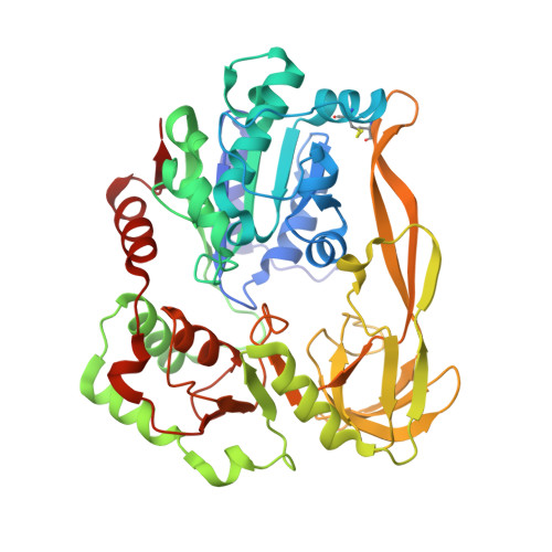
Crystal Structures of the Bspif1 Helicase Reveal that a Major Movement of the 2B SH3 Domain is Required for DNA Unwinding

Chen, W.-F.Dai, Y.-X.Duan, X.-L.Liu, N.-N.Shi, W.Li, N.Li, M.Dou, S.-X.Dong, Y.-H.Rety, S.Xi, X.-G.

(2016) Nucleic Acids Res 44: 2949

  • DOI: https://doi.org/10.1093/nar/gkw033
  • Primary Citation of Related Structures:  
    5FTB, 5FTC, 5FTD, 5FTE, 5FTF

  • PubMed Abstract: 

    Pif1 helicases are ubiquitous members of the SF1B family and are essential for maintaining genome stability. It was speculated that Pif1-specific motifs may fold in specific structures, conferring distinct activities upon it. Here, we report the crystal structures of the Pif1 helicase from Bacteroides spp with and without adenosine triphosphate (ATP) analog/ssDNA. BsPif1 shares structural similarities with RecD2 and Dda helicases but has specific features in the 1B and 2B domains. The highly conserved Pif1 family specific sequence motif interacts with and constraints a putative pin-loop in domain 1B in a precise conformation. More importantly, we found that the 2B domain which contains a specific extended hairpin undergoes a significant rotation and/or movement upon ATP and DNA binding, which is absolutely required for DNA unwinding. We therefore propose a mechanism for DNA unwinding in which the 2B domain plays a predominant role. The fact that the conformational change regulates Pif1 activity may provide insight into the puzzling observation that Pif1 becomes highly processive during break-induced replication in association with Polδ, while the isolated Pif1 has low processivity.


  • Organizational Affiliation

    College of Life Sciences, Northwest A&F University, Yangling, Shaanxi 712100, China.


Macromolecules
Find similar proteins by:  (by identity cutoff)  |  3D Structure
Entity ID: 1
MoleculeChains Sequence LengthOrganismDetailsImage
TPR DOMAIN PROTEIN433Bacteroides sp. 3_1_23Mutation(s): 2 
EC: 3.6.4.12
UniProt
Find proteins for D7K0H3 (Bacteroides sp. 3_1_23)
Explore D7K0H3 
Go to UniProtKB:  D7K0H3
Entity Groups  
Sequence Clusters30% Identity50% Identity70% Identity90% Identity95% Identity100% Identity
UniProt GroupD7K0H3
Sequence Annotations
Expand
  • Reference Sequence
Small Molecules
Ligands 1 Unique
IDChains Name / Formula / InChI Key2D Diagram3D Interactions
ADP
Query on ADP

Download Ideal Coordinates CCD File 
B [auth A]ADENOSINE-5'-DIPHOSPHATE
C10 H15 N5 O10 P2
XTWYTFMLZFPYCI-KQYNXXCUSA-N
Experimental Data & Validation

Experimental Data

  • Method: X-RAY DIFFRACTION
  • Resolution: 2.41 Å
  • R-Value Free: 0.300 
  • R-Value Work: 0.213 
  • R-Value Observed: 0.217 
  • Space Group: P 21 21 21
Unit Cell:
Length ( Å )Angle ( ˚ )
a = 67.708α = 90
b = 73.482β = 90
c = 108.648γ = 90
Software Package:
Software NamePurpose
PHENIXrefinement
XDSdata reduction
XDSdata scaling
PHASERphasing

Structure Validation

View Full Validation Report


Ligand Structure Quality Assessment 


Entry History 

Deposition Data

Revision History  (Full details and data files)

  • Version 1.0: 2016-02-03
    Type: Initial release
  • Version 1.1: 2016-03-02
    Changes: Database references
  • Version 1.2: 2016-04-20
    Changes: Database references
  • Version 2.0: 2019-10-23
    Changes: Atomic model, Data collection, Other
  • Version 2.1: 2024-01-10
    Changes: Data collection, Database references, Derived calculations, Refinement description