5FR3

X-ray crystal structure of aggregation-resistant protective antigen of Bacillus anthracis (mutant S559L T576E)


Experimental Data Snapshot

  • Method: X-RAY DIFFRACTION
  • Resolution: 1.94 Å
  • R-Value Free: 0.227 
  • R-Value Work: 0.178 
  • R-Value Observed: 0.180 

wwPDB Validation   3D Report Full Report


This is version 1.3 of the entry. See complete history


Literature

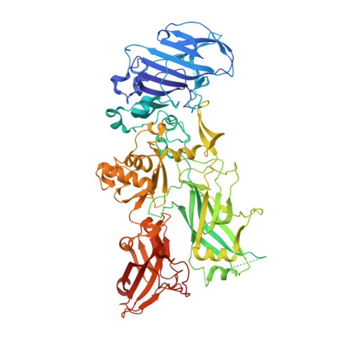
Structural Hot Spots for the Solubility of Globular Proteins

Ganesan, A.Siekierska, A.Beerten, J.Brams, M.Van Durme, J.De Baets, G.Van Der Kant, R.Gallardo, R.Ramakers, M.Langenberg, T.Wilkinson, H.De Smet, F.Ulens, C.Rousseau, F.Schymkowitz, J.

(2016) Nat Commun 7: 10816

  • DOI: https://doi.org/10.1038/ncomms10816
  • Primary Citation of Related Structures:  
    5FR3

  • PubMed Abstract: 

    Natural selection shapes protein solubility to physiological requirements and recombinant applications that require higher protein concentrations are often problematic. This raises the question whether the solubility of natural protein sequences can be improved. We here show an anti-correlation between the number of aggregation prone regions (APRs) in a protein sequence and its solubility, suggesting that mutational suppression of APRs provides a simple strategy to increase protein solubility. We show that mutations at specific positions within a protein structure can act as APR suppressors without affecting protein stability. These hot spots for protein solubility are both structure and sequence dependent but can be computationally predicted. We demonstrate this by reducing the aggregation of human α-galactosidase and protective antigen of Bacillus anthracis through mutation. Our results indicate that many proteins possess hot spots allowing to adapt protein solubility independently of structure and function.


  • Organizational Affiliation

    VIB Switch Laboratory, Flanders Institute for Biotechnology (VIB), 3000 Leuven, Belgium.


Macromolecules
Find similar proteins by:  (by identity cutoff)  |  3D Structure
Entity ID: 1
MoleculeChains Sequence LengthOrganismDetailsImage
PROTECTIVE ANTIGEN722Bacillus anthracisMutation(s): 2 
UniProt
Find proteins for P13423 (Bacillus anthracis)
Explore P13423 
Go to UniProtKB:  P13423
Entity Groups  
Sequence Clusters30% Identity50% Identity70% Identity90% Identity95% Identity100% Identity
UniProt GroupP13423
Sequence Annotations
Expand
  • Reference Sequence
Experimental Data & Validation

Experimental Data

  • Method: X-RAY DIFFRACTION
  • Resolution: 1.94 Å
  • R-Value Free: 0.227 
  • R-Value Work: 0.178 
  • R-Value Observed: 0.180 
  • Space Group: P 21 21 21
Unit Cell:
Length ( Å )Angle ( ˚ )
a = 84.547α = 90
b = 94.714β = 90
c = 100.627γ = 90
Software Package:
Software NamePurpose
PHENIXrefinement
XDSdata reduction
SCALAdata scaling
PHASERphasing

Structure Validation

View Full Validation Report


Entry History 

Revision History  (Full details and data files)

  • Version 1.0: 2016-01-27
    Type: Initial release
  • Version 1.1: 2016-03-02
    Changes: Database references, Structure summary
  • Version 1.2: 2016-03-09
    Changes: Database references
  • Version 1.3: 2024-01-10
    Changes: Data collection, Database references, Derived calculations, Other, Refinement description