5FOH

Crystal structure of the catalytic domain of NcLPMO9A


Experimental Data Snapshot

  • Method: X-RAY DIFFRACTION
  • Resolution: 1.60 Å
  • R-Value Free: 0.178 
  • R-Value Work: 0.154 
  • R-Value Observed: 0.155 

wwPDB Validation   3D Report Full Report


Ligand Structure Quality Assessment 


This is version 1.3 of the entry. See complete history


Literature

Comparison of three seemingly similar lytic polysaccharide monooxygenases fromNeurospora crassasuggests different roles in plant biomass degradation.

Petrovic, D.M.Varnai, A.Dimarogona, M.Mathiesen, G.Sandgren, M.Westereng, B.Eijsink, V.G.H.

(2019) J Biol Chem 

  • DOI: https://doi.org/10.1074/jbc.RA119.008196
  • Primary Citation of Related Structures:  
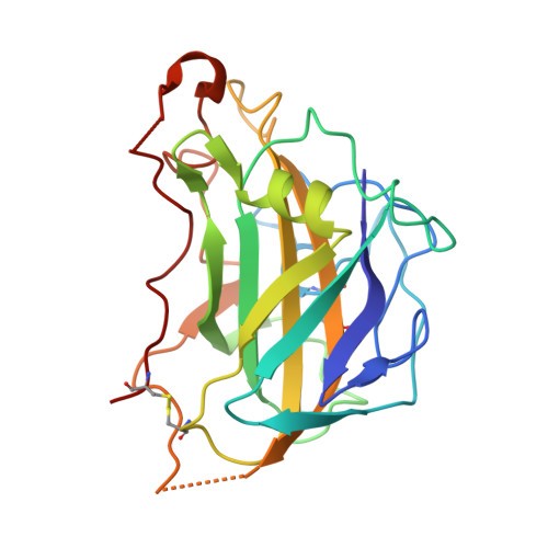
    5FOH

  • PubMed Abstract: 

    Many fungi produce multiple lytic polysaccharide monooxygenases (LPMOs) with seemingly similar functions, but the biological reason for this multiplicity remains unknown. To address this question, here we carried out comparative structural and functional characterizations of three cellulose-active C4-oxidizing family AA9 LPMOs from the fungus Neurospora crassa , Nc LPMO9A (NCU02240), Nc LPMO9C (NCU02916), and Nc LPMO9D (NCU01050). We solved the three-dimensional structure of copper-bound Nc LPMO9A at 1.6-Å resolution and found that Nc LPMO9A and Nc LPMO9C, containing a CBM1 carbohydrate-binding module, bind cellulose more strongly and were less susceptible to inactivation than Nc LPMO9D, which lacks a CBM. All three LPMOs were active on tamarind xyloglucan and konjac glucomannan, generating similar products but clearly differing in activity levels. Importantly, in some cases, the addition of phosphoric acid-swollen cellulose (PASC) had a major effect on activity: Nc LPMO9A was active on xyloglucan only in the presence of PASC, and PASC enhanced Nc LPMO9D activity on glucomannan. Interestingly, the three enzymes also exhibited large differences in their interactions with enzymatic electron donors, which could reflect that they are optimized to act with different reducing partners. All three enzymes efficiently used H 2 O 2 as a cosubstrate, yielding product profiles identical to those obtained in O 2 -driven reactions with PASC, xyloglucan, or glucomannan. Our results indicate that seemingly similar LPMOs act preferentially on different types of copolymeric substructures in the plant cell wall, possibly because these LPMOs are functionally adapted to distinct niches differing in the types of available reductants.


  • Organizational Affiliation

    Faculty of Chemistry, Biotechnology and Food Science, Norwegian University of Life Sciences (NMBU), 1432 Ås, Norway.


Macromolecules
Find similar proteins by:  (by identity cutoff)  |  3D Structure
Entity ID: 1
MoleculeChains Sequence LengthOrganismDetailsImage
POLYSACCHARIDE MONOOXYGENASE223Neurospora crassa OR74AMutation(s): 0 
UniProt
Find proteins for Q7S439 (Neurospora crassa (strain ATCC 24698 / 74-OR23-1A / CBS 708.71 / DSM 1257 / FGSC 987))
Explore Q7S439 
Go to UniProtKB:  Q7S439
Entity Groups  
Sequence Clusters30% Identity50% Identity70% Identity90% Identity95% Identity100% Identity
UniProt GroupQ7S439
Sequence Annotations
Expand
  • Reference Sequence
Small Molecules
Ligands 4 Unique
IDChains Name / Formula / InChI Key2D Diagram3D Interactions
MAN
Query on MAN

Download Ideal Coordinates CCD File 
B [auth A],
C [auth A]
alpha-D-mannopyranose
C6 H12 O6
WQZGKKKJIJFFOK-PQMKYFCFSA-N
SO4
Query on SO4

Download Ideal Coordinates CCD File 
E [auth A],
F [auth A],
G [auth A]
SULFATE ION
O4 S
QAOWNCQODCNURD-UHFFFAOYSA-L
CU
Query on CU

Download Ideal Coordinates CCD File 
D [auth A]COPPER (II) ION
Cu
JPVYNHNXODAKFH-UHFFFAOYSA-N
LI
Query on LI

Download Ideal Coordinates CCD File 
H [auth A]LITHIUM ION
Li
HBBGRARXTFLTSG-UHFFFAOYSA-N
Experimental Data & Validation

Experimental Data

  • Method: X-RAY DIFFRACTION
  • Resolution: 1.60 Å
  • R-Value Free: 0.178 
  • R-Value Work: 0.154 
  • R-Value Observed: 0.155 
  • Space Group: P 32 2 1
Unit Cell:
Length ( Å )Angle ( ˚ )
a = 80.419α = 90
b = 80.419β = 90
c = 57.864γ = 120
Software Package:
Software NamePurpose
REFMACrefinement
XDSdata reduction
Aimlessdata scaling
PHASERphasing

Structure Validation

View Full Validation Report


Ligand Structure Quality Assessment 


Entry History 

Deposition Data

Revision History  (Full details and data files)

  • Version 1.0: 2016-12-07
    Type: Initial release
  • Version 1.1: 2019-09-04
    Changes: Data collection, Database references, Derived calculations
  • Version 1.2: 2020-07-29
    Type: Remediation
    Reason: Carbohydrate remediation
    Changes: Data collection, Derived calculations, Other, Structure summary
  • Version 1.3: 2024-01-10
    Changes: Data collection, Database references, Refinement description, Structure summary