5FKV

cryo-EM structure of the E. coli replicative DNA polymerase complex bound to DNA (DNA polymerase III alpha, beta, epsilon, tau complex)


Experimental Data Snapshot

  • Method: ELECTRON MICROSCOPY
  • Resolution: 8.04 Å
  • Aggregation State: PARTICLE 
  • Reconstruction Method: SINGLE PARTICLE 

wwPDB Validation   3D Report Full Report


This is version 1.5 of the entry. See complete history


Literature

cryo-EM structures of theE. colireplicative DNA polymerase reveal its dynamic interactions with the DNA sliding clamp, exonuclease andtau.

Fernandez-Leiro, R.Conrad, J.Scheres, S.H.Lamers, M.H.

(2015) Elife 4

  • DOI: https://doi.org/10.7554/eLife.11134
  • Primary Citation of Related Structures:  
    5FKU, 5FKV, 5FKW

  • PubMed Abstract: 

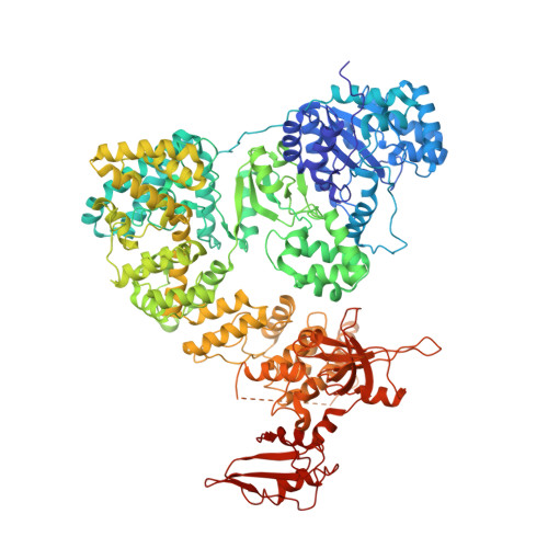
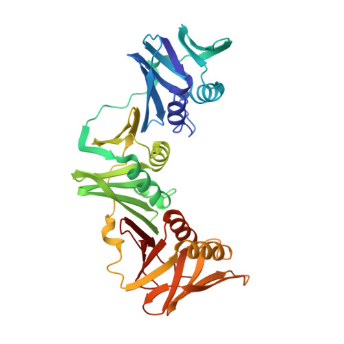
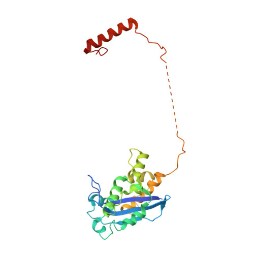
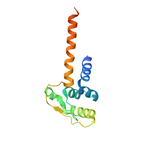
    The replicative DNA polymerase PolIIIα from Escherichia coli is a uniquely fast and processive enzyme. For its activity it relies on the DNA sliding clamp β, the proofreading exonuclease ε and the C-terminal domain of the clamp loader subunit τ. Due to the dynamic nature of the four-protein complex it has long been refractory to structural characterization. Here we present the 8 Å resolution cryo-electron microscopy structures of DNA-bound and DNA-free states of the PolIII-clamp-exonuclease-τ c complex. The structures show how the polymerase is tethered to the DNA through multiple contacts with the clamp and exonuclease. A novel contact between the polymerase and clamp is made in the DNA bound state, facilitated by a large movement of the polymerase tail domain and τ c . These structures provide crucial insights into the organization of the catalytic core of the replisome and form an important step towards determining the structure of the complete holoenzyme.


  • Organizational Affiliation

    MRC Laboratory of Molecular Biology, Cambridge, United Kingdom.


Macromolecules

Find similar proteins by:  (by identity cutoff)  |  3D Structure
Entity ID: 1
MoleculeChains Sequence LengthOrganismDetailsImage
DNA POLYMERASE III SUBUNIT ALPHA1,160Escherichia coli K-12Mutation(s): 2 
EC: 2.7.7.7
UniProt
Find proteins for P10443 (Escherichia coli (strain K12))
Explore P10443 
Go to UniProtKB:  P10443
Entity Groups  
Sequence Clusters30% Identity50% Identity70% Identity90% Identity95% Identity100% Identity
UniProt GroupP10443
Sequence Annotations
Expand
  • Reference Sequence
Find similar proteins by:  (by identity cutoff)  |  3D Structure
Entity ID: 2
MoleculeChains Sequence LengthOrganismDetailsImage
DNA POLYMERASE III BETA
B, C
366Escherichia coli K-12Mutation(s): 0 
EC: 2.7.7.7
UniProt
Find proteins for P0A988 (Escherichia coli (strain K12))
Explore P0A988 
Go to UniProtKB:  P0A988
Entity Groups  
Sequence Clusters30% Identity50% Identity70% Identity90% Identity95% Identity100% Identity
UniProt GroupP0A988
Sequence Annotations
Expand
  • Reference Sequence
Find similar proteins by:  (by identity cutoff)  |  3D Structure
Entity ID: 3
MoleculeChains Sequence LengthOrganismDetailsImage
DNA POLYMERASE III EPSILON243Escherichia coli K-12Mutation(s): 4 
EC: 2.7.7.7
UniProt
Find proteins for P03007 (Escherichia coli (strain K12))
Explore P03007 
Go to UniProtKB:  P03007
Entity Groups  
Sequence Clusters30% Identity50% Identity70% Identity90% Identity95% Identity100% Identity
UniProt GroupP03007
Sequence Annotations
Expand
  • Reference Sequence
Find similar proteins by:  (by identity cutoff)  |  3D Structure
Entity ID: 4
MoleculeChains Sequence LengthOrganismDetailsImage
DNA POLYMERASE III TAU144Escherichia coli K-12Mutation(s): 0 
EC: 2.7.7.7
UniProt
Find proteins for P06710 (Escherichia coli (strain K12))
Explore P06710 
Go to UniProtKB:  P06710
Entity Groups  
Sequence Clusters30% Identity50% Identity70% Identity90% Identity95% Identity100% Identity
UniProt GroupP06710
Sequence Annotations
Expand
  • Reference Sequence

Find similar nucleic acids by:  Sequence   |   3D Structure  

Entity ID: 5
MoleculeChains LengthOrganismImage
PRIMER-TEMPLATE DUPLEX DNAF [auth P]25synthetic construct
Sequence Annotations
Expand
  • Reference Sequence
Find similar nucleic acids by:  (by identity cutoff)  |  3D Structure
Entity ID: 6
MoleculeChains LengthOrganismImage
PRIMER-TEMPLATE DUPLEX DNAG [auth T]29synthetic construct
Sequence Annotations
Expand
  • Reference Sequence
Experimental Data & Validation

Experimental Data

  • Method: ELECTRON MICROSCOPY
  • Resolution: 8.04 Å
  • Aggregation State: PARTICLE 
  • Reconstruction Method: SINGLE PARTICLE 

Structure Validation

View Full Validation Report


Entry History 

Deposition Data

Revision History  (Full details and data files)

  • Version 1.0: 2015-11-25
    Type: Initial release
  • Version 1.1: 2016-12-21
    Changes: Database references
  • Version 1.2: 2017-04-19
    Changes: Other
  • Version 1.3: 2018-06-20
    Changes: Advisory, Data collection, Derived calculations
  • Version 1.4: 2018-10-03
    Changes: Advisory, Data collection, Derived calculations
  • Version 1.5: 2019-02-27
    Changes: Data collection, Database references