Structural and functional characterization of the alanine racemase from Streptomyces coelicolor A3(2).
Tassoni, R., van der Aart, L.T., Ubbink, M., van Wezel, G.P., Pannu, N.S.(2017) Biochem Biophys Res Commun 483: 122-128
- PubMed: 28042035
- DOI: https://doi.org/10.1016/j.bbrc.2016.12.183
- Primary Citation of Related Structures:
5FAC, 5FAG, 5FAJ - PubMed Abstract:
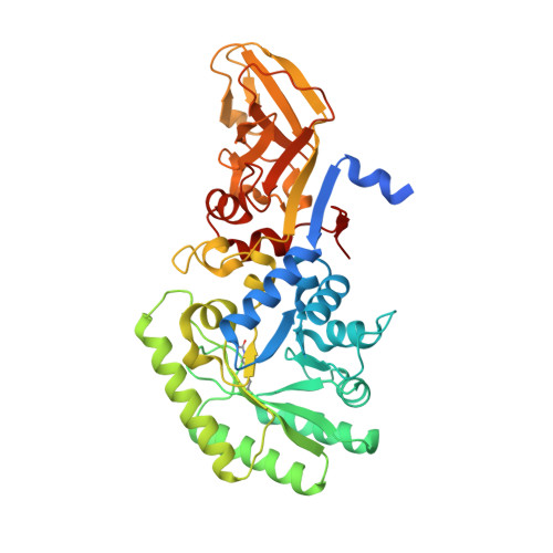
The conversion of l-alanine (L-Ala) into d-alanine (D-Ala) in bacteria is performed by pyridoxal phosphate-dependent enzymes called alanine racemases. D-Ala is an essential component of the bacterial peptidoglycan and hence required for survival. The Gram-positive bacterium Streptomyces coelicolor has at least one alanine racemase encoded by alr. Here, we describe an alr deletion mutant of S. coelicolor which depends on D-Ala for growth and shows increased sensitivity to the antibiotic d-cycloserine (DCS). The crystal structure of the alanine racemase (Alr) was solved with and without the inhibitors DCS or propionate, at 1.64 Å and 1.51 Å resolution, respectively. The crystal structures revealed that Alr is a homodimer with residues from both monomers contributing to the active site. The dimeric state of the enzyme in solution was confirmed by gel filtration chromatography, with and without L-Ala or d-cycloserine. The activity of the enzyme was 66 ± 3 U mg -1 for the racemization of L- to D-Ala, and 104 ± 7 U mg -1 for the opposite direction. Comparison of Alr from S. coelicolor with orthologous enzymes from other bacteria, including the closely related d-cycloserine-resistant Alr from S. lavendulae, strongly suggests that structural features such as the hinge angle or the surface area between the monomers do not contribute to d-cycloserine resistance, and the molecular basis for resistance therefore remains elusive.
Organizational Affiliation:
Leiden Institute of Chemistry, Leiden University, Gorlaeus Laboratories, Einsteinweg 55, 2333 CC Leiden, The Netherlands.