5ET1

Crystal structure of Myo3b-ARB1 in complex with Espin1-AR


Experimental Data Snapshot

  • Method: X-RAY DIFFRACTION
  • Resolution: 1.65 Å
  • R-Value Free: 0.191 
  • R-Value Work: 0.169 
  • R-Value Observed: 0.170 

wwPDB Validation   3D Report Full Report


This is version 1.1 of the entry. See complete history


Literature

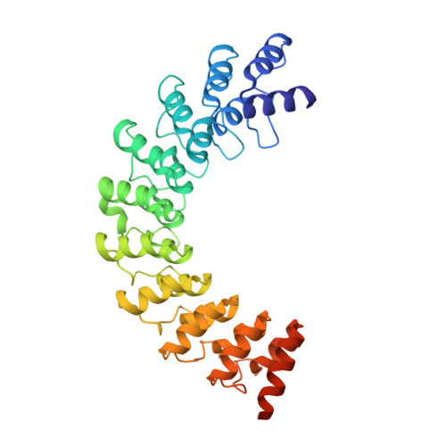
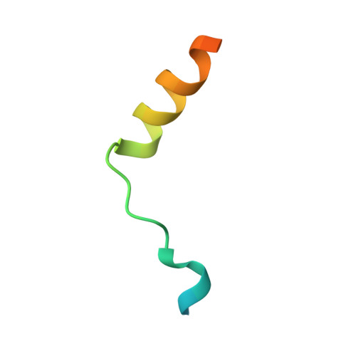
Myosin III-mediated cross-linking and stimulation of actin bundling activity of Espin

Liu, H.Li, J.Raval, M.H.Yao, N.Deng, X.Lu, Q.Nie, S.Feng, W.Wan, J.Yengo, C.M.Liu, W.Zhang, M.

(2016) Elife 5

  • DOI: https://doi.org/10.7554/eLife.12856
  • Primary Citation of Related Structures:  
    5ET0, 5ET1

  • PubMed Abstract: 

    Class III myosins (Myo3) and actin-bundling protein Espin play critical roles in regulating the development and maintenance of stereocilia in vertebrate hair cells, and their defects cause hereditary hearing impairments. Myo3 interacts with Espin1 through its tail homology I motif (THDI), however it is not clear how Myo3 specifically acts through Espin1 to regulate the actin bundle assembly and stabilization. Here we discover that Myo3 THDI contains a pair of repeat sequences capable of independently and strongly binding to the ankyrin repeats of Espin1, revealing an unexpected Myo3-mediated cross-linking mechanism of Espin1. The structures of Myo3 in complex with Espin1 not only elucidate the mechanism of the binding, but also reveal a Myo3-induced release of Espin1 auto-inhibition mechanism. We also provide evidence that Myo3-mediated cross-linking can further promote actin fiber bundling activity of Espin1.


  • Organizational Affiliation

    Shenzhen Key Laboratory for Neuronal Structural Biology, Biomedical Research Institute, Shenzhen Peking University-The Hong Kong University of Science and Technology Medical Center, Shenzhen, China.


Macromolecules
Find similar proteins by:  (by identity cutoff)  |  3D Structure
Entity ID: 1
MoleculeChains Sequence LengthOrganismDetailsImage
Espin
A, B
353Mus musculusMutation(s): 0 
Gene Names: Espn
UniProt & NIH Common Fund Data Resources
Find proteins for Q9ET47 (Mus musculus)
Explore Q9ET47 
Go to UniProtKB:  Q9ET47
IMPC:  MGI:1861630
Entity Groups  
Sequence Clusters30% Identity50% Identity70% Identity90% Identity95% Identity100% Identity
UniProt GroupQ9ET47
Sequence Annotations
Expand
  • Reference Sequence
Find similar proteins by:  (by identity cutoff)  |  3D Structure
Entity ID: 2
MoleculeChains Sequence LengthOrganismDetailsImage
Myosin-IIIb
C, D
36Mus musculusMutation(s): 0 
Gene Names: Myo3b
UniProt & NIH Common Fund Data Resources
Find proteins for Q1EG27 (Mus musculus)
Explore Q1EG27 
Go to UniProtKB:  Q1EG27
IMPC:  MGI:2448580
Entity Groups  
Sequence Clusters30% Identity50% Identity70% Identity90% Identity95% Identity100% Identity
UniProt GroupQ1EG27
Sequence Annotations
Expand
  • Reference Sequence
Experimental Data & Validation

Experimental Data

  • Method: X-RAY DIFFRACTION
  • Resolution: 1.65 Å
  • R-Value Free: 0.191 
  • R-Value Work: 0.169 
  • R-Value Observed: 0.170 
  • Space Group: P 1 21 1
Unit Cell:
Length ( Å )Angle ( ˚ )
a = 72.74α = 90
b = 71.142β = 96.88
c = 76.883γ = 90
Software Package:
Software NamePurpose
PHENIXrefinement
HKL-2000data reduction
HKL-2000data scaling
PHASERphasing

Structure Validation

View Full Validation Report


Entry History & Funding Information

Deposition Data


Funding OrganizationLocationGrant Number
Research Grant CouncilHong Kong--

Revision History  (Full details and data files)

  • Version 1.0: 2016-02-03
    Type: Initial release
  • Version 1.1: 2023-11-08
    Changes: Data collection, Database references, Derived calculations, Refinement description