5ESD

Crystal Structure of M. tuberculosis MenD bound to ThDP and Mn2+


Experimental Data Snapshot

  • Method: X-RAY DIFFRACTION
  • Resolution: 2.25 Å
  • R-Value Free: 0.235 
  • R-Value Work: 0.210 
  • R-Value Observed: 0.212 

wwPDB Validation   3D Report Full Report


Ligand Structure Quality Assessment 


This is version 2.1 of the entry. See complete history


Literature

Structural Views along the Mycobacterium tuberculosis MenD Reaction Pathway Illuminate Key Aspects of Thiamin Diphosphate-Dependent Enzyme Mechanisms.

Jirgis, E.N.Bashiri, G.Bulloch, E.M.Johnston, J.M.Baker, E.N.

(2016) Structure 24: 1167-1177

  • DOI: https://doi.org/10.1016/j.str.2016.04.018
  • Primary Citation of Related Structures:  
    5ERX, 5ERY, 5ESD, 5ESO, 5ESS, 5ESU

  • PubMed Abstract: 

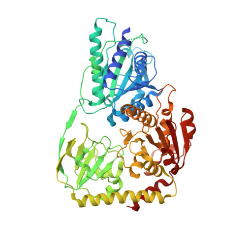
    Menaquinone (MQ) is an essential component of the respiratory chains of many pathogenic organisms, including Mycobacterium tuberculosis (Mtb). The first committed step in MQ biosynthesis is catalyzed by 2-succinyl-5-enolpyruvyl-6-hydroxy-3-cyclohexadiene-1-carboxylate synthase (MenD), a thiamin diphosphate (ThDP)-dependent enzyme. Catalysis proceeds through two covalent intermediates as the substrates 2-oxoglutarate and isochorismate are successively added to the cofactor before final cleavage of the product. We have determined a series of crystal structures of Mtb-MenD that map the binding of both substrates, visualizing each step in the MenD catalytic cycle, including both intermediates. ThDP binding induces a marked asymmetry between the coupled active sites of each dimer, and possible mechanisms of communication can be identified. The crystal structures also reveal conformational features of the two intermediates that facilitate reaction but prevent premature product release. These data fully map chemical space to inform early-stage drug discovery targeting MenD.


  • Organizational Affiliation

    Maurice Wilkins Centre for Molecular Biodiscovery, School of Biological Sciences, University of Auckland, Private Bag 92019, Auckland 1010, New Zealand.


Macromolecules
Find similar proteins by:  (by identity cutoff)  |  3D Structure
Entity ID: 1
MoleculeChains Sequence LengthOrganismDetailsImage
2-succinyl-5-enolpyruvyl-6-hydroxy-3-cyclohexene-1-carboxylate synthase
A, B, C, D
574Mycobacterium tuberculosis H37RvMutation(s): 0 
Gene Names: menDRv0555
EC: 2.2.1.9
UniProt
Find proteins for P9WK11 (Mycobacterium tuberculosis (strain ATCC 25618 / H37Rv))
Explore P9WK11 
Go to UniProtKB:  P9WK11
Entity Groups  
Sequence Clusters30% Identity50% Identity70% Identity90% Identity95% Identity100% Identity
UniProt GroupP9WK11
Sequence Annotations
Expand
  • Reference Sequence
Experimental Data & Validation

Experimental Data

  • Method: X-RAY DIFFRACTION
  • Resolution: 2.25 Å
  • R-Value Free: 0.235 
  • R-Value Work: 0.210 
  • R-Value Observed: 0.212 
  • Space Group: P 21 21 21
Unit Cell:
Length ( Å )Angle ( ˚ )
a = 98.625α = 90
b = 138.657β = 90
c = 165.386γ = 90
Software Package:
Software NamePurpose
Aimlessdata scaling
PHENIXrefinement
PDB_EXTRACTdata extraction
XDSdata scaling
REFMACrefinement
PHASERphasing

Structure Validation

View Full Validation Report


Ligand Structure Quality Assessment 


Entry History 

Deposition Data

Revision History  (Full details and data files)

  • Version 1.0: 2016-06-22
    Type: Initial release
  • Version 1.1: 2016-07-06
    Changes: Database references
  • Version 1.2: 2016-07-20
    Changes: Other
  • Version 1.3: 2017-11-22
    Changes: Data collection, Derived calculations, Refinement description
  • Version 1.4: 2018-01-17
    Changes: Author supporting evidence
  • Version 2.0: 2021-08-04
    Changes: Atomic model, Data collection, Derived calculations, Non-polymer description, Structure summary
  • Version 2.1: 2024-03-06
    Changes: Data collection, Database references