5ERM

Crystal structure of cyclization domain of Phomopsis amygdali fusicoccadiene synthase complexed with magnesium ions and pamidronate


Experimental Data Snapshot

  • Method: X-RAY DIFFRACTION
  • Resolution: 2.30 Å
  • R-Value Free: 0.206 
  • R-Value Work: 0.178 
  • R-Value Observed: 0.182 

wwPDB Validation   3D Report Full Report


Ligand Structure Quality Assessment 


This is version 1.4 of the entry. See complete history


Literature

Structure and Function of Fusicoccadiene Synthase, a Hexameric Bifunctional Diterpene Synthase.

Chen, M.Chou, W.K.Toyomasu, T.Cane, D.E.Christianson, D.W.

(2016) ACS Chem Biol 11: 889-899

  • DOI: https://doi.org/10.1021/acschembio.5b00960
  • Primary Citation of Related Structures:  
    5ER8, 5ERM, 5ERN, 5ERO

  • PubMed Abstract: 

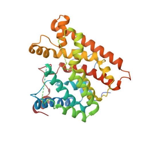
    Fusicoccin A is a diterpene glucoside phytotoxin generated by the fungal pathogen Phomopsis amygdali that causes the plant disease constriction canker, first discovered in New Jersey peach orchards in the 1930s. Fusicoccin A is also an emerging new lead in cancer chemotherapy. The hydrocarbon precursor of fusicoccin A is the tricyclic diterpene fusicoccadiene, which is generated by a bifunctional terpenoid synthase. Here, we report X-ray crystal structures of the individual catalytic domains of fusicoccadiene synthase: the C-terminal domain is a chain elongation enzyme that generates geranylgeranyl diphosphate, and the N-terminal domain catalyzes the cyclization of geranylgeranyl diphosphate to form fusicoccadiene. Crystal structures of each domain complexed with bisphosphonate substrate analogues suggest that three metal ions and three positively charged amino acid side chains trigger substrate ionization in each active site. While in vitro incubations reveal that the cyclase domain can utilize farnesyl diphosphate and geranyl diphosphate as surrogate substrates, these shorter isoprenoid diphosphates are mainly converted into acyclic alcohol or hydrocarbon products. Gel filtration chromatography and analytical ultracentrifugation experiments indicate that full-length fusicoccadiene synthase adopts hexameric quaternary structure, and small-angle X-ray scattering data yield a well-defined molecular envelope illustrating a plausible model for hexamer assembly.


  • Organizational Affiliation

    Roy and Diana Vagelos Laboratories, Department of Chemistry, University of Pennsylvania , Philadelphia, Pennsylvania 19104-6323, United States.


Macromolecules
Find similar proteins by:  (by identity cutoff)  |  3D Structure
Entity ID: 1
MoleculeChains Sequence LengthOrganismDetailsImage
Fusicoccadiene synthase
A, B
363Diaporthe amygdaliMutation(s): 0 
Gene Names: PaFS
EC: 4.2.3.43
UniProt
Find proteins for A2PZA5 (Phomopsis amygdali)
Explore A2PZA5 
Go to UniProtKB:  A2PZA5
Entity Groups  
Sequence Clusters30% Identity50% Identity70% Identity90% Identity95% Identity100% Identity
UniProt GroupA2PZA5
Sequence Annotations
Expand
  • Reference Sequence
Small Molecules
Ligands 3 Unique
IDChains Name / Formula / InChI Key2D Diagram3D Interactions
210
Query on 210

Download Ideal Coordinates CCD File 
C [auth A],
K [auth B]
PAMIDRONATE
C3 H11 N O7 P2
WRUUGTRCQOWXEG-UHFFFAOYSA-N
CL
Query on CL

Download Ideal Coordinates CCD File 
D [auth A],
L [auth B]
CHLORIDE ION
Cl
VEXZGXHMUGYJMC-UHFFFAOYSA-M
MG
Query on MG

Download Ideal Coordinates CCD File 
E [auth A]
F [auth A]
G [auth A]
H [auth B]
I [auth B]
E [auth A],
F [auth A],
G [auth A],
H [auth B],
I [auth B],
J [auth B]
MAGNESIUM ION
Mg
JLVVSXFLKOJNIY-UHFFFAOYSA-N
Experimental Data & Validation

Experimental Data

  • Method: X-RAY DIFFRACTION
  • Resolution: 2.30 Å
  • R-Value Free: 0.206 
  • R-Value Work: 0.178 
  • R-Value Observed: 0.182 
  • Space Group: P 32 2 1
Unit Cell:
Length ( Å )Angle ( ˚ )
a = 143.295α = 90
b = 143.295β = 90
c = 118.261γ = 120
Software Package:
Software NamePurpose
PHENIXrefinement
HKL-2000data reduction
HKL-2000data scaling
PHASERphasing

Structure Validation

View Full Validation Report


Ligand Structure Quality Assessment 


Entry History & Funding Information

Deposition Data


Funding OrganizationLocationGrant Number
National Institutes of Health/National Institute of General Medical Sciences (NIH/NIGMS)United StatesGM56838

Revision History  (Full details and data files)

  • Version 1.0: 2016-01-20
    Type: Initial release
  • Version 1.1: 2016-04-27
    Changes: Database references
  • Version 1.2: 2017-09-20
    Changes: Author supporting evidence, Derived calculations
  • Version 1.3: 2019-12-25
    Changes: Author supporting evidence
  • Version 1.4: 2024-03-06
    Changes: Data collection, Database references, Derived calculations