5EEM

Crystal structure of Danio rerio histone deacetylase 6 catalytic domain 2


Experimental Data Snapshot

  • Method: X-RAY DIFFRACTION
  • Resolution: 2.00 Å
  • R-Value Free: 0.273 
  • R-Value Work: 0.224 
  • R-Value Observed: 0.226 

wwPDB Validation   3D Report Full Report


This is version 1.3 of the entry. See complete history


Literature

Histone deacetylase 6 structure and molecular basis of catalysis and inhibition.

Hai, Y.Christianson, D.W.

(2016) Nat Chem Biol 12: 741-747

  • DOI: https://doi.org/10.1038/nchembio.2134
  • Primary Citation of Related Structures:  
    5EDU, 5EEF, 5EEI, 5EEK, 5EEM, 5EEN, 5EF7, 5EF8, 5EFB, 5EFG, 5EFH, 5EFJ, 5EFK, 5EFN

  • PubMed Abstract: 

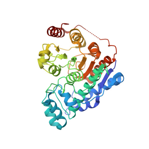
    Histone deacetylase 6 (HDAC6) is a critical target for drug design because of its role in oncogenic transformation and cancer metastasis, and is unique among all histone deacetylases in that it contains tandem catalytic domains designated CD1 and CD2. We now report the crystal structures of CD2 from Homo sapiens HDAC6 and of CD1 and CD2 from Danio rerio HDAC6. We correlated these structures with activity measurements using 13 different substrates. The catalytic activity of CD2 from both species exhibited broad substrate specificity, whereas that of CD1 was highly specific for substrates bearing C-terminal acetyllysine residues. Crystal structures of substrate complexes yielded unprecedented snapshots of the catalytic mechanism. Additionally, crystal structures of complexes with eight different inhibitors, including belinostat and panobinostat (currently used in cancer chemotherapy), the macrocyclic tetrapeptide HC toxin, and the HDAC6-specific inhibitor N-hydroxy-4-(2-((2-hydroxyethyl)(phenyl)amino)-2-oxoethyl)benzamide, revealed surprising new insight regarding changes in Zn(2+) coordination and isozyme-specific inhibition.


  • Organizational Affiliation

    Roy and Diana Vagelos Laboratories, Department of Chemistry, University of Pennsylvania, Philadelphia, Pennsylvania, USA.


Macromolecules
Find similar proteins by:  (by identity cutoff)  |  3D Structure
Entity ID: 1
MoleculeChains Sequence LengthOrganismDetailsImage
Hdac6 proteinA [auth B],
B [auth A]
364Danio rerioMutation(s): 0 
Gene Names: hdac6
EC: 3.5.1.98
UniProt
Find proteins for F8W4B7 (Danio rerio)
Explore F8W4B7 
Go to UniProtKB:  F8W4B7
Entity Groups  
Sequence Clusters30% Identity50% Identity70% Identity90% Identity95% Identity100% Identity
UniProt GroupF8W4B7
Sequence Annotations
Expand
  • Reference Sequence
Small Molecules
Ligands 4 Unique
IDChains Name / Formula / InChI Key2D Diagram3D Interactions
ZN
Query on ZN

Download Ideal Coordinates CCD File 
C [auth B],
H [auth A]
ZINC ION
Zn
PTFCDOFLOPIGGS-UHFFFAOYSA-N
FMT
Query on FMT

Download Ideal Coordinates CCD File 
G [auth B]FORMIC ACID
C H2 O2
BDAGIHXWWSANSR-UHFFFAOYSA-N
K
Query on K

Download Ideal Coordinates CCD File 
D [auth B],
E [auth B],
I [auth A],
J [auth A]
POTASSIUM ION
K
NPYPAHLBTDXSSS-UHFFFAOYSA-N
NA
Query on NA

Download Ideal Coordinates CCD File 
F [auth B]SODIUM ION
Na
FKNQFGJONOIPTF-UHFFFAOYSA-N
Experimental Data & Validation

Experimental Data

  • Method: X-RAY DIFFRACTION
  • Resolution: 2.00 Å
  • R-Value Free: 0.273 
  • R-Value Work: 0.224 
  • R-Value Observed: 0.226 
  • Space Group: P 1
Unit Cell:
Length ( Å )Angle ( ˚ )
a = 48.485α = 73.58
b = 55.4β = 89.84
c = 74.183γ = 82.64
Software Package:
Software NamePurpose
PHENIXrefinement
HKL-2000data reduction
HKL-2000data scaling
PHASERphasing

Structure Validation

View Full Validation Report


Entry History 

Deposition Data

Revision History  (Full details and data files)

  • Version 1.0: 2016-07-27
    Type: Initial release
  • Version 1.1: 2016-08-10
    Changes: Database references
  • Version 1.2: 2016-09-07
    Changes: Database references
  • Version 1.3: 2023-09-27
    Changes: Data collection, Database references, Derived calculations, Refinement description