5DZC

Crystal structure of the cGMP-dependent protein kinase PKG from Plasmodium Vivax - AMPPNP bound


Experimental Data Snapshot

  • Method: X-RAY DIFFRACTION
  • Resolution: 2.30 Å
  • R-Value Free: 0.255 
  • R-Value Work: 0.221 
  • R-Value Observed: 0.223 

wwPDB Validation   3D Report Full Report


Ligand Structure Quality Assessment 


This is version 1.4 of the entry. See complete history


Literature

Structures of the cGMP-dependent protein kinase in malaria parasites reveal a unique structural relay mechanism for activation.

El Bakkouri, M.Kouidmi, I.Wernimont, A.K.Amani, M.Hutchinson, A.Loppnau, P.Kim, J.J.Flueck, C.Walker, J.R.Seitova, A.Senisterra, G.Kakihara, Y.Kim, C.Blackman, M.J.Calmettes, C.Baker, D.A.Hui, R.

(2019) Proc Natl Acad Sci U S A 116: 14164-14173

  • DOI: https://doi.org/10.1073/pnas.1905558116
  • Primary Citation of Related Structures:  
    5DYK, 5DYL, 5DZC, 5E16

  • PubMed Abstract: 

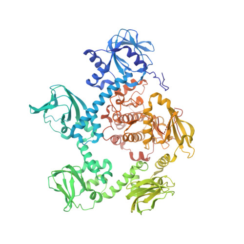
    The cyclic guanosine-3',5'-monophosphate (cGMP)-dependent protein kinase (PKG) was identified >25 y ago; however, efforts to obtain a structure of the entire PKG enzyme or catalytic domain from any species have failed. In malaria parasites, cooperative activation of PKG triggers crucial developmental transitions throughout the complex life cycle. We have determined the cGMP-free crystallographic structures of PKG from Plasmodium falciparum and Plasmodium vivax , revealing how key structural components, including an N-terminal autoinhibitory segment (AIS), four predicted cyclic nucleotide-binding domains (CNBs), and a kinase domain (KD), are arranged when the enzyme is inactive. The four CNBs and the KD are in a pentagonal configuration, with the AIS docked in the substrate site of the KD in a swapped-domain dimeric arrangement. We show that although the protein is predominantly a monomer (the dimer is unlikely to be representative of the physiological form), the binding of the AIS is necessary to keep Plasmodium PKG inactive. A major feature is a helix serving the dual role of the N-terminal helix of the KD as well as the capping helix of the neighboring CNB. A network of connecting helices between neighboring CNBs contributes to maintaining the kinase in its inactive conformation. We propose a scheme in which cooperative binding of cGMP, beginning at the CNB closest to the KD, transmits conformational changes around the pentagonal molecule in a structural relay mechanism, enabling PKG to orchestrate rapid, highly regulated developmental switches in response to dynamic modulation of cGMP levels in the parasite.


  • Organizational Affiliation

    Structural Genomics Consortium, University of Toronto, Toronto, ON M5G 1L7, Canada.


Macromolecules
Find similar proteins by:  (by identity cutoff)  |  3D Structure
Entity ID: 1
MoleculeChains Sequence LengthOrganismDetailsImage
cGMP-dependent protein kinase, putative847Plasmodium vivax Sal-1Mutation(s): 0 
Gene Names: PVX_084705
UniProt
Find proteins for A5K0N4 (Plasmodium vivax (strain Salvador I))
Explore A5K0N4 
Go to UniProtKB:  A5K0N4
Entity Groups  
Sequence Clusters30% Identity50% Identity70% Identity90% Identity95% Identity100% Identity
UniProt GroupA5K0N4
Sequence Annotations
Expand
  • Reference Sequence
Small Molecules
Ligands 4 Unique
IDChains Name / Formula / InChI Key2D Diagram3D Interactions
ANP
Query on ANP

Download Ideal Coordinates CCD File 
B [auth A]PHOSPHOAMINOPHOSPHONIC ACID-ADENYLATE ESTER
C10 H17 N6 O12 P3
PVKSNHVPLWYQGJ-KQYNXXCUSA-N
CL
Query on CL

Download Ideal Coordinates CCD File 
D [auth A],
E [auth A],
F [auth A]
CHLORIDE ION
Cl
VEXZGXHMUGYJMC-UHFFFAOYSA-M
NA
Query on NA

Download Ideal Coordinates CCD File 
G [auth A]SODIUM ION
Na
FKNQFGJONOIPTF-UHFFFAOYSA-N
UNX
Query on UNX

Download Ideal Coordinates CCD File 
C [auth A]
H [auth A]
I [auth A]
J [auth A]
K [auth A]
C [auth A],
H [auth A],
I [auth A],
J [auth A],
K [auth A],
L [auth A],
M [auth A]
UNKNOWN ATOM OR ION
X
Experimental Data & Validation

Experimental Data

  • Method: X-RAY DIFFRACTION
  • Resolution: 2.30 Å
  • R-Value Free: 0.255 
  • R-Value Work: 0.221 
  • R-Value Observed: 0.223 
  • Space Group: C 1 2 1
Unit Cell:
Length ( Å )Angle ( ˚ )
a = 193.111α = 90
b = 117.906β = 95.08
c = 67.731γ = 90
Software Package:
Software NamePurpose
HKL-3000data collection
REFMACrefinement
PDB_EXTRACTdata extraction
HKL-3000data reduction
REFMACphasing

Structure Validation

View Full Validation Report


Ligand Structure Quality Assessment 


Entry History 

Revision History  (Full details and data files)

  • Version 1.0: 2015-10-14
    Type: Initial release
  • Version 1.1: 2018-01-24
    Changes: Database references, Derived calculations, Structure summary
  • Version 1.2: 2019-07-03
    Changes: Data collection, Database references, Structure summary
  • Version 1.3: 2019-11-13
    Changes: Database references
  • Version 1.4: 2023-09-27
    Changes: Data collection, Database references, Refinement description