5DKX

Crystal structure of glucosidase II alpha subunit (Tris-bound from)


Experimental Data Snapshot

  • Method: X-RAY DIFFRACTION
  • Resolution: 1.40 Å
  • R-Value Free: 0.175 
  • R-Value Work: 0.154 
  • R-Value Observed: 0.155 

wwPDB Validation   3D Report Full Report


This is version 1.3 of the entry. See complete history


Literature

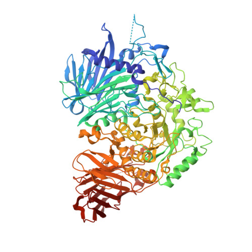
Structural basis for two-step glucose trimming by glucosidase II involved in ER glycoprotein quality control.

Satoh, T.Toshimori, T.Yan, G.Yamaguchi, T.Kato, K.

(2016) Sci Rep 6: 20575-20575

  • DOI: https://doi.org/10.1038/srep20575
  • Primary Citation of Related Structures:  
    5DKX, 5DKY

  • PubMed Abstract: 

    The endoplasmic reticulum (ER) has a sophisticated protein quality control system for the efficient folding of newly synthesized proteins. In this system, a variety of N-linked oligosaccharides displayed on proteins serve as signals recognized by series of intracellular lectins. Glucosidase II catalyzes two-step hydrolysis at α1,3-linked glucose-glucose and glucose-mannose residues of high-mannose-type glycans to generate a quality control protein tag that is transiently expressed on glycoproteins and recognized by ER chaperones. Here we determined the crystal structures of the catalytic α subunit of glucosidase II (GIIα) complexed with two different glucosyl ligands containing the scissile bonds of first- and second-step reactions. Our structural data revealed that the nonreducing terminal disaccharide moieties of the two kinds of substrates can be accommodated in a gourd-shaped bilocular pocket, thereby providing a structural basis for substrate-binding specificity in the two-step deglucosylation catalyzed by this enzyme.


  • Organizational Affiliation

    Graduate School of Pharmaceutical Sciences, Nagoya City University, 3-1 Tanabe-dori, Mizuho-ku, Nagoya 467-8603, Japan.


Macromolecules
Find similar proteins by:  (by identity cutoff)  |  3D Structure
Entity ID: 1
MoleculeChains Sequence LengthOrganismDetailsImage
Alpha glucosidase-like protein951Thermochaetoides thermophila DSM 1495Mutation(s): 0 
Gene Names: CTHT_0064960
UniProt
Find proteins for G0SG42 (Chaetomium thermophilum (strain DSM 1495 / CBS 144.50 / IMI 039719))
Explore G0SG42 
Go to UniProtKB:  G0SG42
Entity Groups  
Sequence Clusters30% Identity50% Identity70% Identity90% Identity95% Identity100% Identity
UniProt GroupG0SG42
Sequence Annotations
Expand
  • Reference Sequence
Experimental Data & Validation

Experimental Data

  • Method: X-RAY DIFFRACTION
  • Resolution: 1.40 Å
  • R-Value Free: 0.175 
  • R-Value Work: 0.154 
  • R-Value Observed: 0.155 
  • Space Group: H 3 2
Unit Cell:
Length ( Å )Angle ( ˚ )
a = 189.026α = 90
b = 189.026β = 90
c = 157.181γ = 120
Software Package:
Software NamePurpose
REFMACrefinement
DENZOdata reduction
SCALEPACKdata scaling
PHENIXphasing

Structure Validation

View Full Validation Report


Entry History & Funding Information

Deposition Data


Funding OrganizationLocationGrant Number
JSPS/MEXTJapan24770102, 25121730 to T.S. and 15H02491, 25102008, 24249002 to K.K.
PRESTO/JSTJapan13417569 to T.S.

Revision History  (Full details and data files)

  • Version 1.0: 2016-01-27
    Type: Initial release
  • Version 1.1: 2016-02-17
    Changes: Database references
  • Version 1.2: 2020-02-19
    Changes: Data collection, Derived calculations
  • Version 1.3: 2024-03-20
    Changes: Data collection, Database references