5DHZ

HIV-1 Rev NTD dimers with variable crossing angles


Experimental Data Snapshot

  • Method: X-RAY DIFFRACTION
  • Resolution: 4.30 Å
  • R-Value Free: 0.324 
  • R-Value Work: 0.328 

wwPDB Validation   3D Report Full Report


This is version 1.4 of the entry. See complete history


Literature

The Structure of HIV-1 Rev Filaments Suggests a Bilateral Model for Rev-RRE Assembly.

DiMattia, M.A.Watts, N.R.Cheng, N.Huang, R.Heymann, J.B.Grimes, J.M.Wingfield, P.T.Stuart, D.I.Steven, A.C.

(2016) Structure 24: 1068-1080

  • DOI: https://doi.org/10.1016/j.str.2016.04.015
  • Primary Citation of Related Structures:  
    5DHV, 5DHX, 5DHY, 5DHZ

  • PubMed Abstract: 

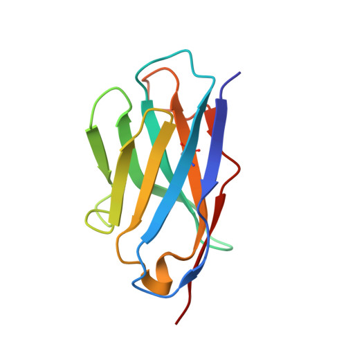
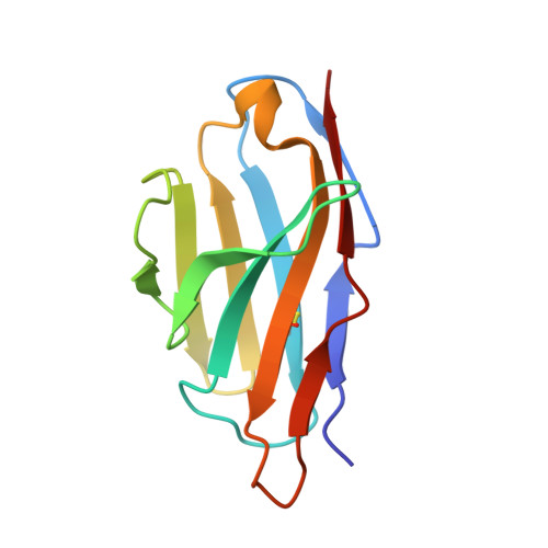
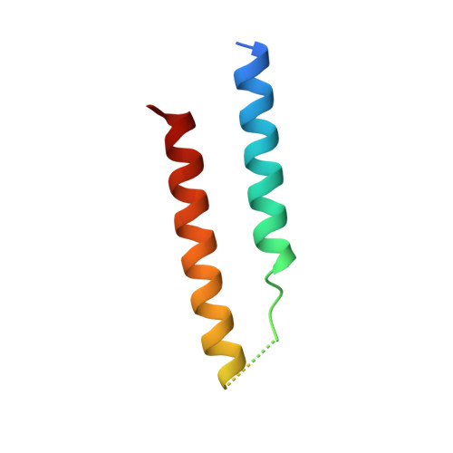
    HIV-1 Rev protein mediates the nuclear export of viral RNA genomes. To do so, Rev oligomerizes cooperatively onto an RNA motif, the Rev response element (RRE), forming a complex that engages with the host nuclear export machinery. To better understand Rev oligomerization, we determined four crystal structures of Rev N-terminal domain dimers, which show that they can pivot about their dyad axis, giving crossing angles of 90° to 140°. In parallel, we performed cryoelectron microscopy of helical Rev filaments. Filaments vary from 11 to 15 nm in width, reflecting variations in dimer crossing angle. These structures contain additional density, indicating that C-terminal domains become partially ordered in the context of filaments. This conformational variability may be exploited in the assembly of RRE/Rev complexes. Our data also revealed a third interface between Revs, which offers an explanation for how the arrangement of Rev subunits adapts to the "A"-shaped architecture of the RRE in export-active complexes.


  • Organizational Affiliation

    Laboratory of Structural Biology Research, National Institute of Arthritis and Musculoskeletal and Skin Diseases, National Institutes of Health, Bethesda, MD 20892, USA; Division of Structural Biology, Henry Wellcome Building for Genomic Medicine, University of Oxford, Roosevelt Drive, Headington OX3 7BN, UK.


Macromolecules
Find similar proteins by:  (by identity cutoff)  |  3D Structure
Entity ID: 1
MoleculeChains Sequence LengthOrganismDetailsImage
Anti-Rev Antibody Fab single-chain variable fragment, heavy chainA [auth H]117Oryctolagus cuniculusMutation(s): 0 
Entity Groups  
Sequence Clusters30% Identity50% Identity70% Identity90% Identity95% Identity100% Identity
Sequence Annotations
Expand
  • Reference Sequence
Find similar proteins by:  (by identity cutoff)  |  3D Structure
Entity ID: 2
MoleculeChains Sequence LengthOrganismDetailsImage
Anti-Rev Antibody Fab single-chain variable fragment, light chainB [auth L]110Oryctolagus cuniculusMutation(s): 0 
Entity Groups  
Sequence Clusters30% Identity50% Identity70% Identity90% Identity95% Identity100% Identity
Sequence Annotations
Expand
  • Reference Sequence
Find similar proteins by:  (by identity cutoff)  |  3D Structure
Entity ID: 3
MoleculeChains Sequence LengthOrganismDetailsImage
Protein RevC [auth M]65Human immunodeficiency virus 1Mutation(s): 0 
Gene Names: rev
UniProt
Find proteins for P04616 (Human immunodeficiency virus type 1 group M subtype B (isolate BH10))
Explore P04616 
Go to UniProtKB:  P04616
Entity Groups  
Sequence Clusters30% Identity50% Identity70% Identity90% Identity95% Identity100% Identity
UniProt GroupP04616
Sequence Annotations
Expand
  • Reference Sequence
Experimental Data & Validation

Experimental Data

  • Method: X-RAY DIFFRACTION
  • Resolution: 4.30 Å
  • R-Value Free: 0.324 
  • R-Value Work: 0.328 
  • Space Group: P 32 1 2
Unit Cell:
Length ( Å )Angle ( ˚ )
a = 48.54α = 90
b = 48.54β = 90
c = 264.482γ = 120
Software Package:
Software NamePurpose
BUSTERrefinement
HKL-2000data reduction
PHASERphasing

Structure Validation

View Full Validation Report


Entry History & Funding Information

Deposition Data


Funding OrganizationLocationGrant Number
National Institutes of HealthUnited States--
SPINE2COMPLEXESUnited KingdomLSHGST-2006-031220
Medical Research Council (United Kingdom)United KingdomG100099

Revision History  (Full details and data files)

  • Version 1.0: 2016-06-29
    Type: Initial release
  • Version 1.1: 2016-07-20
    Changes: Database references
  • Version 1.2: 2017-06-14
    Changes: Refinement description
  • Version 1.3: 2017-08-02
    Changes: Source and taxonomy
  • Version 1.4: 2017-08-30
    Changes: Author supporting evidence