5DGY

Crystal structure of rhodopsin bound to visual arrestin


Experimental Data Snapshot

  • Method: X-RAY DIFFRACTION
  • Resolution: 7.70 Å
  • R-Value Free: 0.335 
  • R-Value Work: 0.285 
  • R-Value Observed: 0.288 

wwPDB Validation   3D Report Full Report


This is version 1.6 of the entry. See complete history


Literature

X-ray laser diffraction for structure determination of the rhodopsin-arrestin complex.

Zhou, X.E.Gao, X.Barty, A.Kang, Y.He, Y.Liu, W.Ishchenko, A.White, T.A.Yefanov, O.Han, G.W.Xu, Q.de Waal, P.W.Suino-Powell, K.M.Boutet, S.Williams, G.J.Wang, M.Li, D.Caffrey, M.Chapman, H.N.Spence, J.C.Fromme, P.Weierstall, U.Stevens, R.C.Cherezov, V.Melcher, K.Xu, H.E.

(2016) Sci Data 3: 160021-160021

  • DOI: https://doi.org/10.1038/sdata.2016.21
  • Primary Citation of Related Structures:  
    5DGY

  • PubMed Abstract: 

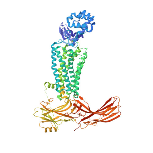
    Serial femtosecond X-ray crystallography (SFX) using an X-ray free electron laser (XFEL) is a recent advancement in structural biology for solving crystal structures of challenging membrane proteins, including G-protein coupled receptors (GPCRs), which often only produce microcrystals. An XFEL delivers highly intense X-ray pulses of femtosecond duration short enough to enable the collection of single diffraction images before significant radiation damage to crystals sets in. Here we report the deposition of the XFEL data and provide further details on crystallization, XFEL data collection and analysis, structure determination, and the validation of the structural model. The rhodopsin-arrestin crystal structure solved with SFX represents the first near-atomic resolution structure of a GPCR-arrestin complex, provides structural insights into understanding of arrestin-mediated GPCR signaling, and demonstrates the great potential of this SFX-XFEL technology for accelerating crystal structure determination of challenging proteins and protein complexes.


  • Organizational Affiliation

    Laboratory of Structural Sciences, Center for Structural Biology and Drug Discovery, Van Andel Research Institute, Grand Rapids, Michigan 49503, USA.


Macromolecules
Find similar proteins by:  (by identity cutoff)  |  3D Structure
Entity ID: 1
MoleculeChains Sequence LengthOrganismDetailsImage
Endolysin,Rhodopsin,S-arrestin
A, B, C, D
906Tequatrovirus T4Homo sapiensMus musculus
This entity is chimeric
Mutation(s): 11 
EC: 3.2.1.17
Membrane Entity: Yes 
UniProt & NIH Common Fund Data Resources
Find proteins for P00720 (Enterobacteria phage T4)
Explore P00720 
Go to UniProtKB:  P00720
Find proteins for P08100 (Homo sapiens)
Explore P08100 
Go to UniProtKB:  P08100
PHAROS:  P08100
GTEx:  ENSG00000163914 
Find proteins for P20443 (Mus musculus)
Explore P20443 
Go to UniProtKB:  P20443
IMPC:  MGI:98227
Entity Groups  
Sequence Clusters30% Identity50% Identity70% Identity90% Identity95% Identity100% Identity
UniProt GroupsP00720P08100P20443
Sequence Annotations
Expand
  • Reference Sequence
Experimental Data & Validation

Experimental Data

Unit Cell:
Length ( Å )Angle ( ˚ )
a = 107.484α = 90
b = 107.258β = 90
c = 460.314γ = 90
Software Package:
Software NamePurpose
XSCALEdata scaling
PHENIXrefinement
PDB_EXTRACTdata extraction
XDSdata reduction
PHASERphasing

Structure Validation

View Full Validation Report


Entry History & Funding Information

Deposition Data


Funding OrganizationLocationGrant Number
National Institutes of Health/National Institute of Diabetes and Digestive and Kidney Disease (NIH/NIDDK)United StatesDK071662
National Institutes of Health/National Cancer Institute (NIH/NCI)United StatesACB-12002
National Institutes of Health/National Institute of General Medical Sciences (NIH/NIGMS)United StatesAGM-12006
Michigan Economic Development Corporation and the Michigan Technology Tri-CorridorUnited States085P1000817

Revision History  (Full details and data files)

  • Version 1.0: 2016-03-23
    Type: Initial release
  • Version 1.1: 2016-04-27
    Changes: Database references
  • Version 1.2: 2017-08-09
    Changes: Database references, Derived calculations
  • Version 1.3: 2017-09-20
    Changes: Author supporting evidence
  • Version 1.4: 2019-12-04
    Changes: Author supporting evidence
  • Version 1.5: 2023-08-16
    Changes: Data collection, Database references
  • Version 1.6: 2023-09-27
    Changes: Refinement description