5D7Z

Crystal structure of glyoxalase I from Zea mays


Experimental Data Snapshot

  • Method: X-RAY DIFFRACTION
  • Resolution: 1.73 Å
  • R-Value Free: 0.202 
  • R-Value Work: 0.166 
  • R-Value Observed: 0.168 

wwPDB Validation   3D Report Full Report


This is version 1.3 of the entry. See complete history


Literature

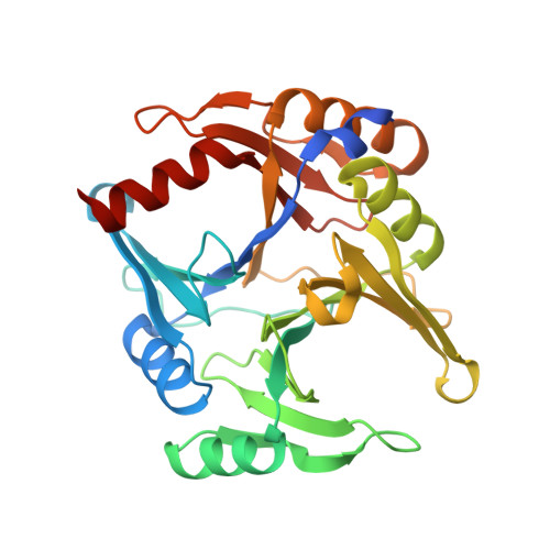
Structure of the novel monomeric glyoxalase I from Zea mays.

Turra, G.L.Agostini, R.B.Fauguel, C.M.Presello, D.A.Andreo, C.S.Gonzalez, J.M.Campos-Bermudez, V.A.

(2015) Acta Crystallogr D Biol Crystallogr 71: 2009-2020

  • DOI: https://doi.org/10.1107/S1399004715015205
  • Primary Citation of Related Structures:  
    5D7Z

  • PubMed Abstract: 

    The glyoxalase system is ubiquitous among all forms of life owing to its central role in relieving the cell from the accumulation of methylglyoxal, a toxic metabolic byproduct. In higher plants, this system is upregulated under diverse metabolic stress conditions, such as in the defence response to infection by pathogenic microorganisms. Despite their proven fundamental role in metabolic stresses, plant glyoxalases have been poorly studied. In this work, glyoxalase I from Zea mays has been characterized both biochemically and structurally, thus reporting the first atomic model of a glyoxalase I available from plants. The results indicate that this enzyme comprises a single polypeptide with two structurally similar domains, giving rise to two lateral concavities, one of which harbours a functional nickel(II)-binding active site. The putative function of the remaining cryptic active site remains to be determined.


  • Organizational Affiliation

    Centro de Estudios Fotosintéticos y Bioquímicos (CEFOBI-CONICET), Universidad Nacional de Rosario, Suipacha 531, S2002LRK Rosario, Argentina.


Macromolecules
Find similar proteins by:  (by identity cutoff)  |  3D Structure
Entity ID: 1
MoleculeChains Sequence LengthOrganismDetailsImage
Lactoylglutathione lyase298Zea maysMutation(s): 0 
Gene Names: Zm.67052ZEAMMB73_635823
EC: 4.4.1.5
UniProt
Find proteins for B6TPH0 (Zea mays)
Explore B6TPH0 
Go to UniProtKB:  B6TPH0
Entity Groups  
Sequence Clusters30% Identity50% Identity70% Identity90% Identity95% Identity100% Identity
UniProt GroupB6TPH0
Sequence Annotations
Expand
  • Reference Sequence
Small Molecules
Ligands 4 Unique
IDChains Name / Formula / InChI Key2D Diagram3D Interactions
2PE
Query on 2PE

Download Ideal Coordinates CCD File 
I [auth A]NONAETHYLENE GLYCOL
C18 H38 O10
YZUUTMGDONTGTN-UHFFFAOYSA-N
NI
Query on NI

Download Ideal Coordinates CCD File 
B [auth A]NICKEL (II) ION
Ni
VEQPNABPJHWNSG-UHFFFAOYSA-N
FMT
Query on FMT

Download Ideal Coordinates CCD File 
C [auth A],
D [auth A],
E [auth A],
F [auth A],
G [auth A]
FORMIC ACID
C H2 O2
BDAGIHXWWSANSR-UHFFFAOYSA-N
NA
Query on NA

Download Ideal Coordinates CCD File 
H [auth A]SODIUM ION
Na
FKNQFGJONOIPTF-UHFFFAOYSA-N
Experimental Data & Validation

Experimental Data

  • Method: X-RAY DIFFRACTION
  • Resolution: 1.73 Å
  • R-Value Free: 0.202 
  • R-Value Work: 0.166 
  • R-Value Observed: 0.168 
  • Space Group: P 63
Unit Cell:
Length ( Å )Angle ( ˚ )
a = 90.69α = 90
b = 90.69β = 90
c = 74.713γ = 120
Software Package:
Software NamePurpose
REFMACrefinement
MOSFLMdata reduction
SCALAdata scaling
PHASERphasing

Structure Validation

View Full Validation Report


Entry History 

Deposition Data

  • Released Date: 2015-09-09 
  • Deposition Author(s): Gonzalez, J.M.
  • This entry supersedes: 4RO6

Revision History  (Full details and data files)

  • Version 1.0: 2015-09-09
    Type: Initial release
  • Version 1.1: 2015-10-14
    Changes: Database references
  • Version 1.2: 2015-10-28
    Changes: Database references
  • Version 1.3: 2024-03-06
    Changes: Data collection, Database references, Derived calculations