5D2R

Inhibitor Bound Cell Shape Determinant Protein Csd4 from Helicobacter pylori


Experimental Data Snapshot

  • Method: X-RAY DIFFRACTION
  • Resolution: 1.90 Å
  • R-Value Free: 0.219 
  • R-Value Work: 0.180 
  • R-Value Observed: 0.182 

wwPDB Validation   3D Report Full Report


Ligand Structure Quality Assessment 


This is version 1.2 of the entry. See complete history


Literature

A Bacterial Cell Shape-Determining Inhibitor.

Liu, Y.Frirdich, E.Taylor, J.A.Chan, A.C.Blair, K.M.Vermeulen, J.Ha, R.Murphy, M.E.Salama, N.R.Gaynor, E.C.Tanner, M.E.

(2016) ACS Chem Biol 11: 981-991

  • DOI: https://doi.org/10.1021/acschembio.5b01039
  • Primary Citation of Related Structures:  
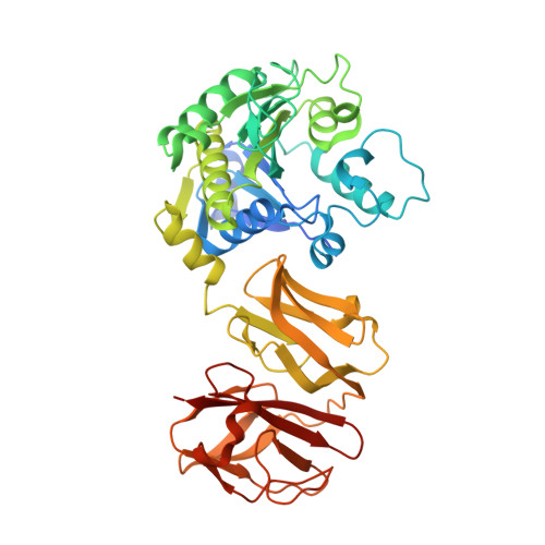
    5D2R

  • PubMed Abstract: 

    Helicobacter pylori and Campylobacter jejuni are human pathogens and causative agents of gastric ulcers/cancer and gastroenteritis, respectively. Recent studies have uncovered a series of proteases that are responsible for maintaining the helical shape of these organisms. The H. pylori metalloprotease Csd4 and its C. jejuni homologue Pgp1 cleave the amide bond between meso-diaminopimelate and iso-d-glutamic acid in truncated peptidoglycan side chains. Deletion of either csd4 or pgp1 results in bacteria with a straight rod phenotype, a reduced ability to move in viscous media, and reduced pathogenicity. In this work, a phosphinic acid-based pseudodipeptide inhibitor was designed to act as a tetrahedral intermediate analog against the Csd4 enzyme. The phosphinic acid was shown to inhibit the cleavage of the alternate substrate, Ac-l-Ala-iso-d-Glu-meso-Dap, with a Ki value of 1.5 μM. Structural analysis of the Csd4-inhibitor complex shows that the phosphinic acid displaces the zinc-bound water and chelates the metal in a bidentate fashion. The phosphinate oxygens also interact with the key acid/base residue, Glu222, and the oxyanion-stabilizing residue, Arg86. The results are consistent with the "promoted-water pathway" mechanism for carboxypeptidase A catalysis. Studies on cultured bacteria showed that the inhibitor causes significant cell straightening when incubated with H. pylori at millimolar concentrations. A diminished, yet observable, effect on the morphology of C. jejuni was also apparent. Cell straightening was more pronounced with an acapsular C. jejuni mutant strain compared to the wild type, suggesting that the capsule impaired inhibitor accessibility. These studies demonstrate that a highly polar compound is capable of crossing the outer membrane and altering cell shape, presumably by inhibiting cell shape determinant proteases. Peptidoglycan proteases acting as cell shape determinants represent novel targets for the development of antimicrobials against these human pathogens.


  • Organizational Affiliation

    Contribution from the Department of Chemistry, University of British Columbia , Vancouver, British Columbia V6T 1Z1, Canada.


Macromolecules
Find similar proteins by:  (by identity cutoff)  |  3D Structure
Entity ID: 1
MoleculeChains Sequence LengthOrganismDetailsImage
Conserved hypothetical secreted protein439Helicobacter pylori G27Mutation(s): 0 
Gene Names: HPG27_353
UniProt
Find proteins for B5ZAD9 (Helicobacter pylori (strain G27))
Explore B5ZAD9 
Go to UniProtKB:  B5ZAD9
Entity Groups  
Sequence Clusters30% Identity50% Identity70% Identity90% Identity95% Identity100% Identity
UniProt GroupB5ZAD9
Sequence Annotations
Expand
  • Reference Sequence
Small Molecules
Ligands 3 Unique
IDChains Name / Formula / InChI Key2D Diagram3D Interactions
56W
Query on 56W

Download Ideal Coordinates CCD File 
X [auth A](2R,6R)-2-{[(R)-[(3R)-3-(acetylamino)-3-carboxypropyl](hydroxy)phosphoryl]methyl}-6-aminoheptanedioic acid
C14 H25 N2 O9 P
AWMPEAXWUINOES-HBNTYKKESA-N
IOD
Query on IOD

Download Ideal Coordinates CCD File 
E [auth A]
F [auth A]
G [auth A]
H [auth A]
I [auth A]
E [auth A],
F [auth A],
G [auth A],
H [auth A],
I [auth A],
J [auth A],
K [auth A],
L [auth A],
M [auth A],
N [auth A],
O [auth A],
P [auth A],
Q [auth A],
R [auth A],
S [auth A],
T [auth A],
U [auth A],
V [auth A],
W [auth A]
IODIDE ION
I
XMBWDFGMSWQBCA-UHFFFAOYSA-M
ZN
Query on ZN

Download Ideal Coordinates CCD File 
B [auth A],
C [auth A],
D [auth A]
ZINC ION
Zn
PTFCDOFLOPIGGS-UHFFFAOYSA-N
Binding Affinity Annotations 
IDSourceBinding Affinity
56W Binding MOAD:  5D2R Ki: 1500 (nM) from 1 assay(s)
Experimental Data & Validation

Experimental Data

  • Method: X-RAY DIFFRACTION
  • Resolution: 1.90 Å
  • R-Value Free: 0.219 
  • R-Value Work: 0.180 
  • R-Value Observed: 0.182 
  • Space Group: P 21 21 21
Unit Cell:
Length ( Å )Angle ( ˚ )
a = 52.84α = 90
b = 66.61β = 90
c = 144.48γ = 90
Software Package:
Software NamePurpose
XSCALEdata scaling
PHENIXrefinement
PDB_EXTRACTdata extraction
XDSdata reduction
PHENIXphasing

Structure Validation

View Full Validation Report


Ligand Structure Quality Assessment 


Entry History 

Deposition Data

Revision History  (Full details and data files)

  • Version 1.0: 2016-01-20
    Type: Initial release
  • Version 1.1: 2016-04-27
    Changes: Database references
  • Version 1.2: 2023-09-27
    Changes: Data collection, Database references, Derived calculations, Refinement description