


Crystal structure of an A-form DNA duplex containing 5-hydroxylmethylcytidine
Sheng, J.To be published.
Experimental Data Snapshot
wwPDB Validation 3D Report Full Report

Find similar nucleic acids by: Sequence | 3D Structure
Entity ID: 1 | |||||
|---|---|---|---|---|---|
| Molecule | Chains | Length | Organism | Image | |
| DNA (5'-R(*G)-D(P*(UMS))-R(P*G)-D(P*T)-R(P*A)-D(P*(5HC))-R(P*AP*C)-3') | 8 | synthetic construct | 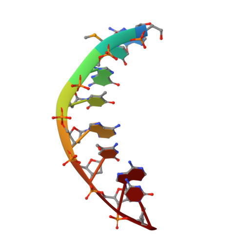 | ||
Sequence AnnotationsExpand | |||||
| |||||
| Length ( Å ) | Angle ( ˚ ) |
|---|---|
| a = 43.05 | α = 90 |
| b = 43.05 | β = 90 |
| c = 23.764 | γ = 90 |
| Software Name | Purpose |
|---|---|
| REFMAC | refinement |
| HKL-2000 | data reduction |
| HKL-2000 | data scaling |
| PHASER | phasing |
RCSB PDB (citation) is hosted by
RCSB PDB is a member of the

