5C9B

Crystal structure of a retropepsin-like aspartic protease from Rickettsia conorii


Experimental Data Snapshot

  • Method: X-RAY DIFFRACTION
  • Resolution: 2.40 Å
  • R-Value Free: 0.262 
  • R-Value Work: 0.199 
  • R-Value Observed: 0.202 

wwPDB Validation   3D Report Full Report


This is version 1.2 of the entry. See complete history


Literature

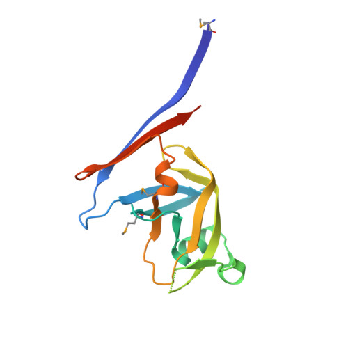
Structure of RC1339/APRc from Rickettsia conorii, a retropepsin-like aspartic protease.

Li, M.Gustchina, A.Cruz, R.Simoes, M.Curto, P.Martinez, J.Faro, C.Simoes, I.Wlodawer, A.

(2015) Acta Crystallogr D Biol Crystallogr 71: 2109-2118

  • DOI: https://doi.org/10.1107/S1399004715013905
  • Primary Citation of Related Structures:  
    5C9B, 5C9F

  • PubMed Abstract: 

    The crystal structures of two constructs of RC1339/APRc from Rickettsia conorii, consisting of either residues 105-231 or 110-231 followed by a His tag, have been determined in three different crystal forms. As predicted, the fold of a monomer of APRc resembles one-half of the mandatory homodimer of retroviral pepsin-like aspartic proteases (retropepsins), but the quaternary structure of the dimer of APRc differs from that of the canonical retropepsins. The observed dimer is most likely an artifact of the expression and/or crystallization conditions since it cannot support the previously reported enzymatic activity of this bacterial aspartic protease. However, the fold of the core of each monomer is very closely related to the fold of retropepsins from a variety of retroviruses and to a single domain of pepsin-like eukaryotic enzymes, and may represent a putative common ancestor of monomeric and dimeric aspartic proteases.


  • Organizational Affiliation

    Protein Structure Section, Macromolecular Crystallography Laboratory, National Cancer Institute, Frederick, MD 21702, USA.


Macromolecules
Find similar proteins by:  (by identity cutoff)  |  3D Structure
Entity ID: 1
MoleculeChains Sequence LengthOrganismDetailsImage
ApRick protease
A, B, C, D
139Rickettsia conoriiMutation(s): 0 
Gene Names: RC1339
EC: 3
UniProt
Find proteins for Q92FY8 (Rickettsia conorii (strain ATCC VR-613 / Malish 7))
Explore Q92FY8 
Go to UniProtKB:  Q92FY8
Entity Groups  
Sequence Clusters30% Identity50% Identity70% Identity90% Identity95% Identity100% Identity
UniProt GroupQ92FY8
Sequence Annotations
Expand
  • Reference Sequence
Small Molecules
Modified Residues  1 Unique
IDChains TypeFormula2D DiagramParent
MSE
Query on MSE
A, B, C, D
L-PEPTIDE LINKINGC5 H11 N O2 SeMET
Experimental Data & Validation

Experimental Data

  • Method: X-RAY DIFFRACTION
  • Resolution: 2.40 Å
  • R-Value Free: 0.262 
  • R-Value Work: 0.199 
  • R-Value Observed: 0.202 
  • Space Group: P 32 2 1
Unit Cell:
Length ( Å )Angle ( ˚ )
a = 101.985α = 90
b = 101.985β = 90
c = 127.063γ = 120
Software Package:
Software NamePurpose
REFMACrefinement
HKL-2000data reduction
SCALEPACKdata scaling
SOLVEphasing

Structure Validation

View Full Validation Report


Entry History 

Deposition Data

Revision History  (Full details and data files)

  • Version 1.0: 2015-10-14
    Type: Initial release
  • Version 1.1: 2018-04-18
    Changes: Data collection, Database references, Derived calculations
  • Version 1.2: 2019-03-13
    Changes: Data collection, Database references