5C3U

Crystal structure of a fungal L-serine ammonia-lyase from Rhizomucor miehei


Experimental Data Snapshot

  • Method: X-RAY DIFFRACTION
  • Resolution: 1.76 Å
  • R-Value Free: 0.209 
  • R-Value Work: 0.186 
  • R-Value Observed: 0.188 

wwPDB Validation   3D Report Full Report


Ligand Structure Quality Assessment 


This is version 1.1 of the entry. See complete history


Literature

Crystal structure and characterization of a novel l-serine ammonia-lyase from Rhizomucor miehei.

Qin, Z.Yan, Q.Ma, Q.Jiang, Z.

(2015) Biochem Biophys Res Commun 466: 431-437

  • DOI: https://doi.org/10.1016/j.bbrc.2015.09.043
  • Primary Citation of Related Structures:  
    5C3U

  • PubMed Abstract: 

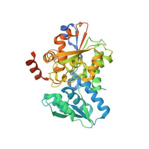
    L-serine ammonia-lyase, as a member of the β-family of pyridoxal-5'-phosphate (PLP) dependent enzymes, catalyzes the conversion of L-serine (L-threonine) to pyruvate (α-ketobutyrate) and ammonia. The crystal structure of L-serine ammonia-lyase from Rhizomucor miehei (RmSDH) was solved at 1.76 Å resolution by X-ray diffraction method. The overall structure of RmSDH had the characteristic β-family PLP dependent enzyme fold. It consisted of two distinct domains, both of which show the typical open twisted α/β structure. A PLP cofactor was located in the crevice between the two domains, which was attached to Lys52 by a Schiff-base linkage. Unique residue substitutions (Gly78, Pro79, Ser146, Ser147 and Thr312) were discovered at the catalytic site of RmSDH by comparison of structures of RmSDH and other reported eukaryotic L-serine ammonia-lyases. Optimal pH and temperature of the purified RmSDH were 7.5 and 40 °C, respectively. It was stable in the pH range of 7.0-9.0 and at temperatures below 40 °C. This is the first crystal structure of a fungal L-serine ammonia-lyase. It will be useful to study the catalytic mechanism of β-elimination enzymes and will provide a basis for further enzyme engineering.


  • Organizational Affiliation

    College of Food Science and Nutritional Engineering, Beijing Advanced Innovation Center of Food Nutrition and Human Health, China Agricultural University, Beijing 100083, China.


Macromolecules
Find similar proteins by:  (by identity cutoff)  |  3D Structure
Entity ID: 1
MoleculeChains Sequence LengthOrganismDetailsImage
L-serine ammonia-lyase366Rhizomucor miehei CAU432Mutation(s): 0 
EC: 4.3.1.17
UniProt
Find proteins for A0A0X1KHE6 (Rhizomucor miehei CAU432)
Explore A0A0X1KHE6 
Go to UniProtKB:  A0A0X1KHE6
Entity Groups  
Sequence Clusters30% Identity50% Identity70% Identity90% Identity95% Identity100% Identity
UniProt GroupA0A0X1KHE6
Sequence Annotations
Expand
  • Reference Sequence
Small Molecules
Ligands 1 Unique
IDChains Name / Formula / InChI Key2D Diagram3D Interactions
PLP
Query on PLP

Download Ideal Coordinates CCD File 
B [auth A]PYRIDOXAL-5'-PHOSPHATE
C8 H10 N O6 P
NGVDGCNFYWLIFO-UHFFFAOYSA-N
Experimental Data & Validation

Experimental Data

  • Method: X-RAY DIFFRACTION
  • Resolution: 1.76 Å
  • R-Value Free: 0.209 
  • R-Value Work: 0.186 
  • R-Value Observed: 0.188 
  • Space Group: C 2 2 21
Unit Cell:
Length ( Å )Angle ( ˚ )
a = 72.98α = 90
b = 72.885β = 90
c = 111.121γ = 90
Software Package:
Software NamePurpose
REFMACrefinement
HKL-2000data reduction
HKL-2000data scaling
Cootmodel building

Structure Validation

View Full Validation Report


Ligand Structure Quality Assessment 


Entry History 

Deposition Data

Revision History  (Full details and data files)

  • Version 1.0: 2015-12-09
    Type: Initial release
  • Version 1.1: 2024-01-10
    Changes: Data collection, Database references, Derived calculations, Refinement description