5BT5

Crystal structure of BRD2 second bromodomain in complex with SGC-CBP30 chemical probe


Experimental Data Snapshot

  • Method: X-RAY DIFFRACTION
  • Resolution: 1.40 Å
  • R-Value Free: 0.182 
  • R-Value Work: 0.160 
  • R-Value Observed: 0.161 

wwPDB Validation   3D Report Full Report


Ligand Structure Quality Assessment 


This is version 1.0 of the entry. See complete history


Literature

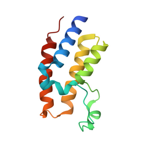
Crystal structure of BRD2 second bromodomain in complex with a 3,5-dimethylisoxazol ligand

Tallant, C.Hay, D.Krojer, T.Nunez-Alonso, G.Picaud, S.Newman, J.A.Fedorov, O.von Delft, F.Arrowsmith, C.H.Edwards, A.M.Bountra, C.Brennan, P.E.Knapp, S.

To be published.

Macromolecules
Find similar proteins by:  (by identity cutoff)  |  3D Structure
Entity ID: 1
MoleculeChains Sequence LengthOrganismDetailsImage
Bromodomain-containing protein 2114Homo sapiensMutation(s): 0 
Gene Names: BRD2KIAA9001RING3
UniProt & NIH Common Fund Data Resources
Find proteins for P25440 (Homo sapiens)
Explore P25440 
Go to UniProtKB:  P25440
PHAROS:  P25440
GTEx:  ENSG00000204256 
Entity Groups  
Sequence Clusters30% Identity50% Identity70% Identity90% Identity95% Identity100% Identity
UniProt GroupP25440
Sequence Annotations
Expand
  • Reference Sequence
Experimental Data & Validation

Experimental Data

  • Method: X-RAY DIFFRACTION
  • Resolution: 1.40 Å
  • R-Value Free: 0.182 
  • R-Value Work: 0.160 
  • R-Value Observed: 0.161 
  • Space Group: P 21 21 2
Unit Cell:
Length ( Å )Angle ( ˚ )
a = 52.761α = 90
b = 71.806β = 90
c = 31.996γ = 90
Software Package:
Software NamePurpose
REFMACrefinement
XDSdata reduction
SCALAdata scaling
PHASERphasing

Structure Validation

View Full Validation Report


Ligand Structure Quality Assessment 


Entry History 

Revision History  (Full details and data files)

  • Version 1.0: 2015-07-01
    Type: Initial release