5B51

Crystal structure of heme binding protein HmuT R242A mutant


Experimental Data Snapshot

  • Method: X-RAY DIFFRACTION
  • Resolution: 1.30 Å
  • R-Value Free: 0.181 
  • R-Value Work: 0.163 
  • R-Value Observed: 0.163 

wwPDB Validation   3D Report Full Report


Ligand Structure Quality Assessment 


This is version 1.2 of the entry. See complete history


Literature

Structural Characterization of Heme Environmental Mutants of CgHmuT that Shuttles Heme Molecules to Heme Transporters

Muraki, N.Kitatsuji, C.Ogura, M.Uchida, T.Ishimori, K.Aono, S.

(2016) Int J Mol Sci 17

  • DOI: https://doi.org/10.3390/ijms17060829
  • Primary Citation of Related Structures:  
    5B4Z, 5B50, 5B51

  • PubMed Abstract: 

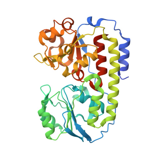
    Corynebacteria contain a heme uptake system encoded in hmuTUV genes, in which HmuT protein acts as a heme binding protein to transport heme to the cognate transporter HmuUV. The crystal structure of HmuT from Corynebacterium glutamicum (CgHmuT) reveals that heme is accommodated in the central cleft with His141 and Tyr240 as the axial ligands and that Tyr240 forms a hydrogen bond with Arg242. In this work, the crystal structures of H141A, Y240A, and R242A mutants were determined to understand the role of these residues for the heme binding of CgHmuT. Overall and heme environmental structures of these mutants were similar to those of the wild type, suggesting that there is little conformational change in the heme-binding cleft during heme transport reaction with binding and the dissociation of heme. A loss of one axial ligand or the hydrogen bonding interaction with Tyr240 resulted in an increase in the redox potential of the heme for CgHmuT to be reduced by dithionite, though the wild type was not reduced under physiological conditions. These results suggest that the heme environmental structure stabilizes the ferric heme binding in CgHmuT, which will be responsible for efficient heme uptake under aerobic conditions where Corynebacteria grow.


  • Organizational Affiliation

    Institute for Molecular Science, National Institutes of Natural Sciences, 5-1 Higashiyama, Myodaiji, Okazaki 444-8787, Japan. nmuraki@ims.ac.jp.


Macromolecules
Find similar proteins by:  (by identity cutoff)  |  3D Structure
Entity ID: 1
MoleculeChains Sequence LengthOrganismDetailsImage
ABC-type transporter, periplasmic component345Corynebacterium glutamicum ATCC 13032Mutation(s): 1 
Gene Names: Cgl0389
UniProt
Find proteins for Q8NTB8 (Corynebacterium glutamicum (strain ATCC 13032 / DSM 20300 / BCRC 11384 / JCM 1318 / LMG 3730 / NCIMB 10025))
Explore Q8NTB8 
Go to UniProtKB:  Q8NTB8
Entity Groups  
Sequence Clusters30% Identity50% Identity70% Identity90% Identity95% Identity100% Identity
UniProt GroupQ8NTB8
Sequence Annotations
Expand
  • Reference Sequence
Small Molecules
Ligands 1 Unique
IDChains Name / Formula / InChI Key2D Diagram3D Interactions
HEM
Query on HEM

Download Ideal Coordinates CCD File 
B [auth A]PROTOPORPHYRIN IX CONTAINING FE
C34 H32 Fe N4 O4
KABFMIBPWCXCRK-RGGAHWMASA-L
Experimental Data & Validation

Experimental Data

  • Method: X-RAY DIFFRACTION
  • Resolution: 1.30 Å
  • R-Value Free: 0.181 
  • R-Value Work: 0.163 
  • R-Value Observed: 0.163 
  • Space Group: P 41 21 2
Unit Cell:
Length ( Å )Angle ( ˚ )
a = 73.06α = 90
b = 73.06β = 90
c = 147.07γ = 90
Software Package:
Software NamePurpose
REFMACrefinement
XDSdata reduction
XSCALEdata scaling
MOLREPphasing

Structure Validation

View Full Validation Report


Ligand Structure Quality Assessment 


Entry History 

Deposition Data

Revision History  (Full details and data files)

  • Version 1.0: 2017-03-01
    Type: Initial release
  • Version 1.1: 2020-02-26
    Changes: Data collection, Derived calculations
  • Version 1.2: 2023-11-08
    Changes: Data collection, Database references, Refinement description