5B2G

Crystal structure of human claudin-4 in complex with C-terminal fragment of Clostridium perfringens enterotoxin


Experimental Data Snapshot

  • Method: X-RAY DIFFRACTION
  • Resolution: 3.50 Å
  • R-Value Free: 0.309 
  • R-Value Work: 0.288 
  • R-Value Observed: 0.289 

wwPDB Validation   3D Report Full Report


This is version 1.4 of the entry. See complete history


Literature

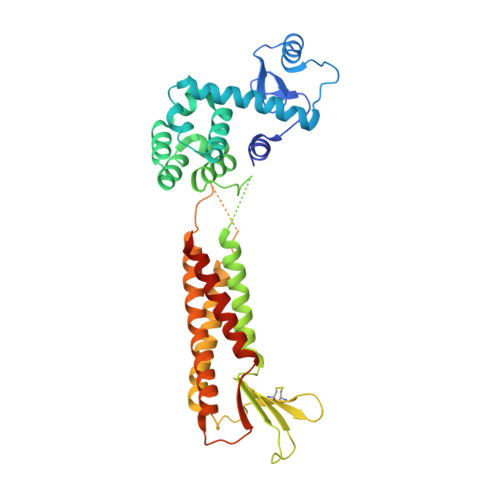
Structural basis for disruption of claudin assembly in tight junctions by an enterotoxin

Shinoda, T.Shinya, N.Ito, K.Ohsawa, N.Terada, T.Hirata, K.Kawano, Y.Yamamoto, M.Kimura-Someya, T.Yokoyama, S.Shirouzu, M.

(2016) Sci Rep 6: 33632-33632

  • DOI: https://doi.org/10.1038/srep33632
  • Primary Citation of Related Structures:  
    5B2G

  • PubMed Abstract: 

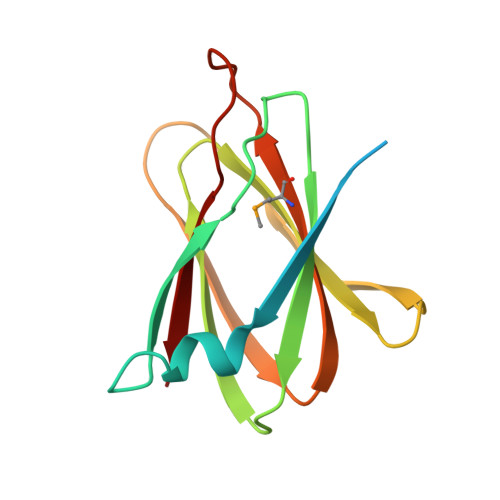
    The food-poisoning bacterium Clostridium perfringens produces an enterotoxin (~35 kDa) that specifically targets human claudin-4, among the 26 human claudin proteins, and causes diarrhea by fluid accumulation in the intestinal cavity. The C-terminal domain of the Clostridium perfringens enterotoxin (C-CPE, ~15 kDa) binds tightly to claudin-4, and disrupts the intestinal tight junction barriers. In this study, we determined the 3.5-Å resolution crystal structure of the cell-free synthesized human claudin-4•C-CPE complex, which is significantly different from the structure of the off-target complex of an engineered C-CPE with mouse claudin-19. The claudin-4•C-CPE complex structure demonstrated the mechanism underlying claudin assembly disruption. A comparison of the present C-CPE-bound structure of claudin-4 with the enterotoxin-free claudin-15 structure revealed sophisticated C-CPE-induced conformation changes of the extracellular segments, induced on the foundation of the rigid four-transmembrane-helix bundle structure. These conformation changes provide a mechanistic model for the disruption of the lateral assembly of claudin molecules. Furthermore, the present novel structural mechanism for selecting a specific member of the claudin family can be used as the foundation to develop novel medically important technologies to selectively regulate the tight junctions formed by claudin family members in different organs.


  • Organizational Affiliation

    RIKEN Systems and Structural Biology Center, 1-7-22 Suehiro-cho, Tsurumi-ku, Yokohama 230-0045, Japan.


Macromolecules
Find similar proteins by:  (by identity cutoff)  |  3D Structure
Entity ID: 1
MoleculeChains Sequence LengthOrganismDetailsImage
Endolysin,Claudin-4
A, C, E, G
364Tequatrovirus T4Homo sapiens
This entity is chimeric
Mutation(s): 4 
EC: 3.2.1.17
Membrane Entity: Yes 
UniProt & NIH Common Fund Data Resources
Find proteins for O14493 (Homo sapiens)
Explore O14493 
Go to UniProtKB:  O14493
PHAROS:  O14493
GTEx:  ENSG00000189143 
Find proteins for P00720 (Enterobacteria phage T4)
Explore P00720 
Go to UniProtKB:  P00720
Entity Groups  
Sequence Clusters30% Identity50% Identity70% Identity90% Identity95% Identity100% Identity
UniProt GroupsP00720O14493
Sequence Annotations
Expand
  • Reference Sequence
Find similar proteins by:  (by identity cutoff)  |  3D Structure
Entity ID: 2
MoleculeChains Sequence LengthOrganismDetailsImage
Heat-labile enterotoxin B chain
B, D, F, H
142Clostridium perfringensMutation(s): 0 
Gene Names: cpe
UniProt
Find proteins for P01558 (Clostridium perfringens)
Explore P01558 
Go to UniProtKB:  P01558
Entity Groups  
Sequence Clusters30% Identity50% Identity70% Identity90% Identity95% Identity100% Identity
UniProt GroupP01558
Sequence Annotations
Expand
  • Reference Sequence
Small Molecules
Modified Residues  1 Unique
IDChains TypeFormula2D DiagramParent
MSE
Query on MSE
B, D, F, H
L-PEPTIDE LINKINGC5 H11 N O2 SeMET
Experimental Data & Validation

Experimental Data

  • Method: X-RAY DIFFRACTION
  • Resolution: 3.50 Å
  • R-Value Free: 0.309 
  • R-Value Work: 0.288 
  • R-Value Observed: 0.289 
  • Space Group: P 43
Unit Cell:
Length ( Å )Angle ( ˚ )
a = 105.92α = 90
b = 105.92β = 90
c = 244.28γ = 90
Software Package:
Software NamePurpose
PHENIXrefinement
XDSdata reduction
XDSdata scaling
PHENIXphasing

Structure Validation

View Full Validation Report


Entry History & Funding Information

Deposition Data


Funding OrganizationLocationGrant Number
Target Proteins Research ProgramJapan--
Platform for Drug Discovery, Information, and Structural Life ScienceJapan--
Grant-in-Aid for Young Scientists (B)Japan25840028

Revision History  (Full details and data files)

  • Version 1.0: 2016-10-05
    Type: Initial release
  • Version 1.1: 2016-10-19
    Changes: Source and taxonomy
  • Version 1.2: 2020-02-26
    Changes: Data collection, Derived calculations
  • Version 1.3: 2023-11-08
    Changes: Data collection, Database references, Refinement description
  • Version 1.4: 2023-11-15
    Changes: Data collection