Unique Flap Conformation in an HIV-1 Protease with High-Level Darunavir Resistance
Nakashima, M., Ode, H., Suzuki, K., Fujino, M., Maejima, M., Kimura, Y., Masaoka, T., Hattori, J., Matsuda, M., Hachiya, A., Yokomaku, Y., Suzuki, A., Watanabe, N., Sugiura, W., Iwatani, Y.(2016) Front Microbiol 7: 61-61
- PubMed: 26870021
- DOI: https://doi.org/10.3389/fmicb.2016.00061
- Primary Citation of Related Structures:
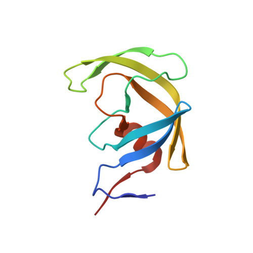
5B18 - PubMed Abstract:
Darunavir (DRV) is one of the most powerful protease inhibitors (PIs) for treating human immunodeficiency virus type-1 (HIV-1) infection and presents a high genetic barrier to the generation of resistant viruses. However, DRV-resistant HIV-1 infrequently emerges from viruses exhibiting resistance to other protease inhibitors. To address this resistance, researchers have gathered genetic information on DRV resistance. In contrast, few structural insights into the mechanism underlying DRV resistance are available. To elucidate this mechanism, we determined the crystal structure of the ligand-free state of a protease with high-level DRV resistance and six DRV resistance-associated mutations (including I47V and I50V), which we generated by in vitro selection. This crystal structure showed a unique curling conformation at the flap regions that was not found in the previously reported ligand-free protease structures. Molecular dynamics simulations indicated that the curled flap conformation altered the flap dynamics. These results suggest that the preference for a unique flap conformation influences DRV binding. These results provide new structural insights into elucidating the molecular mechanism of DRV resistance and aid to develop PIs effective against DRV-resistant viruses.
Organizational Affiliation:
Department of Infectious Diseases and Immunology, Clinical Research Center, National Hospital Organization Nagoya Medical CenterNagoya, Japan; Department of Biotechnology, Nagoya University Graduate School of EngineeringNagoya, Japan.