5AOG

Structure of Sorghum peroxidase


Experimental Data Snapshot

  • Method: X-RAY DIFFRACTION
  • Resolution: 1.27 Å
  • R-Value Free: 0.159 
  • R-Value Work: 0.136 
  • R-Value Observed: 0.137 

wwPDB Validation   3D Report Full Report


Ligand Structure Quality Assessment 


This is version 2.1 of the entry. See complete history


Literature

Structural and Spectroscopic Characterisation of a Heme Peroxidase from Sorghum.

Nnamchi, C.I.Parkin, G.Efimov, I.Basran, J.Kwon, H.Svistunenko, D.A.Agirre, J.Okolo, B.N.Moneke, A.Nwanguma, B.C.Moody, P.C.E.Raven, E.L.

(2016) J Biol Inorg Chem 21: 63

  • DOI: https://doi.org/10.1007/s00775-015-1313-z
  • Primary Citation of Related Structures:  
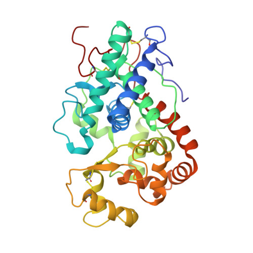
    5AOG

  • PubMed Abstract: 

    A cationic class III peroxidase from Sorghum bicolor was purified to homogeneity. The enzyme contains a high-spin heme, as evidenced by UV-visible spectroscopy and EPR. Steady state oxidation of guaiacol was demonstrated and the enzyme was shown to have higher activity in the presence of calcium ions. A Fe(III)/Fe(II) reduction potential of -266 mV vs NHE was determined. Stopped-flow experiments with H2O2 showed formation of a typical peroxidase Compound I species, which converts to Compound II in the presence of calcium. A crystal structure of the enzyme is reported, the first for a sorghum peroxidase. The structure reveals an active site that is analogous to those for other class I heme peroxidase, and a substrate binding site (assigned as arising from binding of indole-3-acetic acid) at the γ-heme edge. Metal binding sites are observed in the structure on the distal (assigned as a Na(+) ion) and proximal (assigned as a Ca(2+)) sides of the heme, which is consistent with the Ca(2+)-dependence of the steady state and pre-steady state kinetics. It is probably the case that the structural integrity (and, thus, the catalytic activity) of the sorghum enzyme is dependent on metal ion incorporation at these positions.


  • Organizational Affiliation

    Department of Microbiology, University of Nigeria, Nsukka, Nigeria.


Macromolecules
Find similar proteins by:  (by identity cutoff)  |  3D Structure
Entity ID: 1
MoleculeChains Sequence LengthOrganismDetailsImage
CATIONIC PEROXIDASE SPC4307Sorghum bicolorMutation(s): 0 
EC: 1.11.1.7
UniProt
Find proteins for P84516 (Sorghum bicolor)
Explore P84516 
Go to UniProtKB:  P84516
Entity Groups  
Sequence Clusters30% Identity50% Identity70% Identity90% Identity95% Identity100% Identity
UniProt GroupP84516
Sequence Annotations
Expand
  • Reference Sequence
Oligosaccharides

Help

Entity ID: 2
MoleculeChains Length2D Diagram Glycosylation3D Interactions
alpha-L-fucopyranose-(1-3)-[2-acetamido-2-deoxy-beta-D-glucopyranose-(1-4)]2-acetamido-2-deoxy-beta-D-glucopyranose
B
3N-Glycosylation
Glycosylation Resources
GlyTouCan:  G11971MR
GlyCosmos:  G11971MR
GlyGen:  G11971MR
Entity ID: 3
MoleculeChains Length2D Diagram Glycosylation3D Interactions
beta-D-xylopyranose-(1-2)-[alpha-D-mannopyranose-(1-3)]beta-D-mannopyranose-(1-4)-2-acetamido-2-deoxy-beta-D-glucopyranose-(1-4)-[alpha-L-fucopyranose-(1-3)]2-acetamido-2-deoxy-beta-D-glucopyranose
C
6N-Glycosylation
Glycosylation Resources
GlyTouCan:  G68461PT
GlyCosmos:  G68461PT
GlyGen:  G68461PT
Small Molecules
Ligands 5 Unique
IDChains Name / Formula / InChI Key2D Diagram3D Interactions
HEM
Query on HEM

Download Ideal Coordinates CCD File 
D [auth A]PROTOPORPHYRIN IX CONTAINING FE
C34 H32 Fe N4 O4
KABFMIBPWCXCRK-RGGAHWMASA-L
IAC
Query on IAC

Download Ideal Coordinates CCD File 
H [auth A]1H-INDOL-3-YLACETIC ACID
C10 H9 N O2
SEOVTRFCIGRIMH-UHFFFAOYSA-N
GOL
Query on GOL

Download Ideal Coordinates CCD File 
G [auth A]GLYCEROL
C3 H8 O3
PEDCQBHIVMGVHV-UHFFFAOYSA-N
CA
Query on CA

Download Ideal Coordinates CCD File 
E [auth A]CALCIUM ION
Ca
BHPQYMZQTOCNFJ-UHFFFAOYSA-N
NA
Query on NA

Download Ideal Coordinates CCD File 
F [auth A]SODIUM ION
Na
FKNQFGJONOIPTF-UHFFFAOYSA-N
Experimental Data & Validation

Experimental Data

  • Method: X-RAY DIFFRACTION
  • Resolution: 1.27 Å
  • R-Value Free: 0.159 
  • R-Value Work: 0.136 
  • R-Value Observed: 0.137 
  • Space Group: P 31 2 1
Unit Cell:
Length ( Å )Angle ( ˚ )
a = 58.62α = 90
b = 58.62β = 90
c = 208.41γ = 120
Software Package:
Software NamePurpose
REFMACrefinement
XDSdata reduction
Aimlessdata scaling
PHASERphasing

Structure Validation

View Full Validation Report


Ligand Structure Quality Assessment 


Entry History 

Deposition Data

Revision History  (Full details and data files)

  • Version 1.0: 2016-01-13
    Type: Initial release
  • Version 1.1: 2016-03-16
    Changes: Database references
  • Version 2.0: 2020-07-29
    Type: Remediation
    Reason: Carbohydrate remediation
    Changes: Advisory, Atomic model, Data collection, Derived calculations, Other, Structure summary
  • Version 2.1: 2024-01-10
    Changes: Data collection, Database references, Derived calculations, Refinement description, Structure summary