5AHY

Halorhodopsin from Halobacterium salinarum in a new rhombohedral crystal form


Experimental Data Snapshot

  • Method: X-RAY DIFFRACTION
  • Resolution: 2.15 Å
  • R-Value Free: 0.227 
  • R-Value Work: 0.185 
  • R-Value Observed: 0.187 

wwPDB Validation   3D Report Full Report


Ligand Structure Quality Assessment 


This is version 1.6 of the entry. See complete history


Literature

Structure of Halorhodopsin from Halobacterium Salinarum in a New Crystal Form that Imposes Little Restraint on the E-F Loop.

Schreiner, M.Schlesinger, R.Heberle, J.Niemann, H.H.

(2015) J Struct Biol 190: 373

  • DOI: https://doi.org/10.1016/j.jsb.2015.04.010
  • Primary Citation of Related Structures:  
    5AHY, 5AHZ

  • PubMed Abstract: 

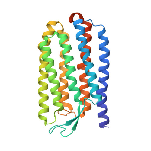
    Halorhodopsin from the halophilic archaeon Halobacterium salinarum is a membrane located light-driven chloride pump. Upon illumination Halorhodopsin undergoes a reversible photocycle initiated by the all-trans to 13-cis isomerization of the covalently bound retinal chromophore. The photocycle consists of several spectroscopically distinct intermediates. The structural basis of the chloride transport mechanism remains elusive, presumably because packing contacts have so far precluded protein conformational changes in the available crystals. With the intention to structurally characterize late photocycle intermediates by X-ray crystallography we crystallized Halorhodopsin in a new crystal form using the vesicle fusion method. In the new crystal form lateral contacts are mediated by helices A and G. Helices E and F that were suggested to perform large movements during the photocycle are almost unrestrained by packing contacts. This feature might permit the displacement of these helices without disrupting the crystal lattice. Therefore, this new crystal form might be an excellent system for the structural characterization of late Halorhodopsin photocycle intermediates by trapping or by time resolved experiments, especially at XFELs.


  • Organizational Affiliation

    Department of Chemistry, Bielefeld University, Universitätsstraße 25, 33615 Bielefeld, Germany.


Macromolecules
Find similar proteins by:  (by identity cutoff)  |  3D Structure
Entity ID: 1
MoleculeChains Sequence LengthOrganismDetailsImage
HALORHODOPSIN261Halobacterium salinarumMutation(s): 0 
Membrane Entity: Yes 
UniProt
Find proteins for B0R2U4 (Halobacterium salinarum (strain ATCC 29341 / DSM 671 / R1))
Explore B0R2U4 
Go to UniProtKB:  B0R2U4
Entity Groups  
Sequence Clusters30% Identity50% Identity70% Identity90% Identity95% Identity100% Identity
UniProt GroupB0R2U4
Sequence Annotations
Expand
  • Reference Sequence
Experimental Data & Validation

Experimental Data

  • Method: X-RAY DIFFRACTION
  • Resolution: 2.15 Å
  • R-Value Free: 0.227 
  • R-Value Work: 0.185 
  • R-Value Observed: 0.187 
  • Space Group: H 3 2
Unit Cell:
Length ( Å )Angle ( ˚ )
a = 103.651α = 90
b = 103.651β = 90
c = 138.588γ = 120
Software Package:
Software NamePurpose
PHENIXrefinement
XDSdata reduction
XSCALEdata scaling
PHASERphasing

Structure Validation

View Full Validation Report


Ligand Structure Quality Assessment 


Entry History 

Deposition Data

Revision History  (Full details and data files)

  • Version 1.0: 2015-04-22
    Type: Initial release
  • Version 1.1: 2015-05-13
    Changes: Database references
  • Version 1.2: 2015-06-17
    Changes: Database references
  • Version 1.3: 2019-05-08
    Changes: Data collection, Derived calculations, Experimental preparation, Other
  • Version 1.4: 2019-09-25
    Changes: Data collection, Experimental preparation, Other
  • Version 1.5: 2020-07-29
    Type: Remediation
    Reason: Carbohydrate remediation
    Changes: Data collection, Derived calculations, Structure summary
  • Version 1.6: 2024-01-10
    Changes: Data collection, Database references, Refinement description, Structure summary