5AHM

IMP-bound form of the DeltaCBS mutant of IMPDH from Pseudomonas aeruginosa


Experimental Data Snapshot

  • Method: X-RAY DIFFRACTION
  • Resolution: 1.74 Å
  • R-Value Free: 0.189 
  • R-Value Work: 0.161 
  • R-Value Observed: 0.162 

wwPDB Validation   3D Report Full Report


Ligand Structure Quality Assessment 


This is version 1.3 of the entry. See complete history


Literature

Crystallographic Studies of Two Variants of Pseudomonas Aeruginosa Impdh with Impaired Allosteric Regulation

Labesse, G.Alexandre, T.Gelin, M.Haouz, A.Munier-Lehmann, H.

(2015) Acta Crystallogr D Biol Crystallogr 71: 1890

  • DOI: https://doi.org/10.1107/S1399004715013115
  • Primary Citation of Related Structures:  
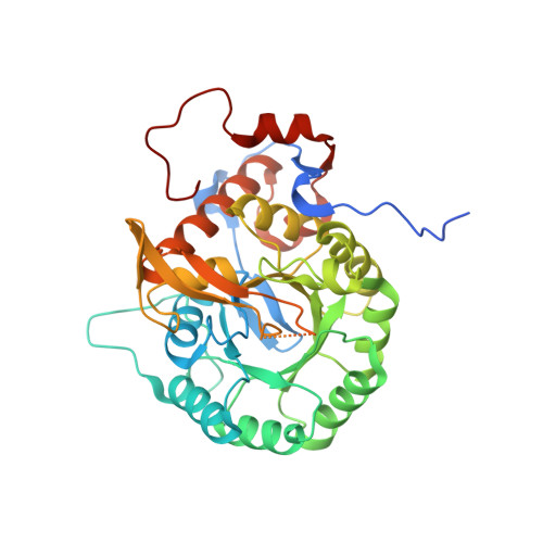
    5AHL, 5AHM, 5AHN

  • PubMed Abstract: 

    Inosine-5'-monophosphate dehydrogenases (IMPDHs), which are the rate-limiting enzymes in guanosine-nucleotide biosynthesis, are important therapeutic targets. Despite in-depth functional and structural characterizations of various IMPDHs, the role of the Bateman domain containing two CBS motifs remains controversial. Their involvement in the allosteric regulation of Pseudomonas aeruginosa IMPDH by Mg-ATP has recently been reported. To better understand the function of IMPDH and the importance of the CBS motifs, the structure of a variant devoid of these modules (ΔCBS) was solved at high resolution in the apo form and in complex with IMP. In addition, a single amino-acid substitution variant, D199N, was also structurally characterized: the mutation corresponds to the autosomal dominant mutant D226N of human IMPDH1, which is responsible for the onset of the retinopathy adRP10. These new structures shed light onto the possible mechanism of regulation of the IMPDH enzymatic activity. In particular, three conserved loops seem to be key players in this regulation as they connect the tetramer-tetramer interface with the active site and show significant modification upon substrate binding.


  • Organizational Affiliation

    CNRS, UMR5048, Université Montpellier 1 et 2, Centre de Biochimie Structurale, 34090 Montpellier, France.


Macromolecules
Find similar proteins by:  (by identity cutoff)  |  3D Structure
Entity ID: 1
MoleculeChains Sequence LengthOrganismDetailsImage
INOSINE-5'-MONOPHOSPHATE DEHYDROGENASE
A, B
400Pseudomonas aeruginosa PAO1Mutation(s): 0 
EC: 1.1.1.205
UniProt
Find proteins for Q9HXM5 (Pseudomonas aeruginosa (strain ATCC 15692 / DSM 22644 / CIP 104116 / JCM 14847 / LMG 12228 / 1C / PRS 101 / PAO1))
Explore Q9HXM5 
Go to UniProtKB:  Q9HXM5
Entity Groups  
Sequence Clusters30% Identity50% Identity70% Identity90% Identity95% Identity100% Identity
UniProt GroupQ9HXM5
Sequence Annotations
Expand
  • Reference Sequence
Experimental Data & Validation

Experimental Data

  • Method: X-RAY DIFFRACTION
  • Resolution: 1.74 Å
  • R-Value Free: 0.189 
  • R-Value Work: 0.161 
  • R-Value Observed: 0.162 
  • Space Group: I 4
Unit Cell:
Length ( Å )Angle ( ˚ )
a = 103.6α = 90
b = 103.6β = 90
c = 158.4γ = 90
Software Package:
Software NamePurpose
PHENIXrefinement
XDSdata reduction
SCALAdata scaling
PHASERphasing

Structure Validation

View Full Validation Report


Ligand Structure Quality Assessment 


Entry History 

Deposition Data

Revision History  (Full details and data files)

  • Version 1.0: 2015-07-15
    Type: Initial release
  • Version 1.1: 2015-09-09
    Changes: Database references
  • Version 1.2: 2015-09-16
    Changes: Database references
  • Version 1.3: 2024-01-10
    Changes: Data collection, Database references, Derived calculations, Other, Refinement description