5A8R

METHYL-COENZYME M REDUCTASE II FROM METHANOTHERMOBACTER MARBURGENSIS AT 2.15 A RESOLUTION


Experimental Data Snapshot

  • Method: X-RAY DIFFRACTION
  • Resolution: 2.15 Å
  • R-Value Free: 0.196 
  • R-Value Work: 0.156 
  • R-Value Observed: 0.158 

wwPDB Validation   3D Report Full Report


Ligand Structure Quality Assessment 


This is version 2.2 of the entry. See complete history


Literature

Didehydroaspartate Modification in Methyl-Coenzyme M Reductase Catalyzing Methane Formation.

Wagner, T.Kahnt, J.Ermler, U.Shima, S.

(2016) Angew Chem Int Ed Engl 55: 10630

  • DOI: https://doi.org/10.1002/anie.201603882
  • Primary Citation of Related Structures:  
    5A0Y, 5A8K, 5A8R, 5A8W

  • PubMed Abstract: 

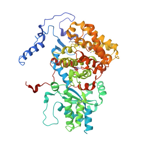
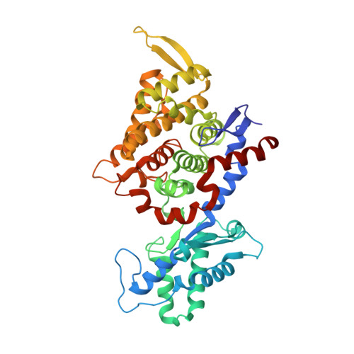
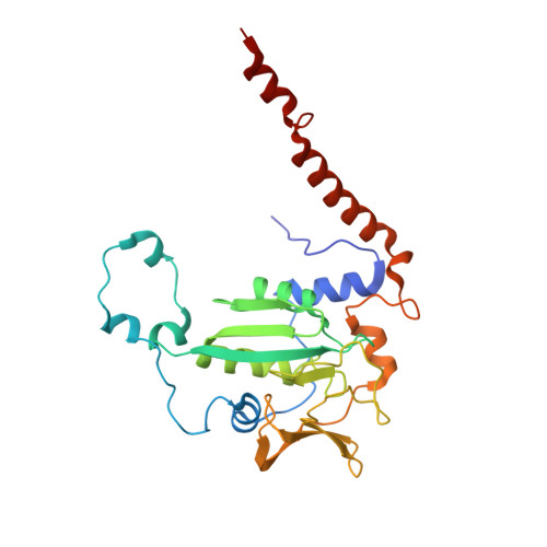
    All methanogenic and methanotrophic archaea known to date contain methyl-coenzyme M reductase (MCR) that catalyzes the reversible reduction of methyl-coenzyme M to methane. This enzyme contains the nickel porphinoid F430 as a prosthetic group and, highly conserved, a thioglycine and four methylated amino acid residues near the active site. We describe herein the presence of a novel post-translationally modified amino acid, didehydroaspartate, adjacent to the thioglycine as revealed by mass spectrometry and high-resolution X-ray crystallography. Upon chemical reduction, the didehydroaspartate residue was converted into aspartate. Didehydroaspartate was found in MCR I and II from Methanothermobacter marburgensis and in MCR of phylogenetically distantly related Methanosarcina barkeri but not in MCR I and II of Methanothermobacter wolfeii, which indicates that didehydroaspartate is dispensable but might have a role in fine-tuning the active site to increase the catalytic efficiency.


  • Organizational Affiliation

    Max-Planck-Institut für terrestrische Mikrobiologie, Karl-von-Frisch-Strasse 10, 35043, Marburg, Germany.


Macromolecules
Find similar proteins by:  (by identity cutoff)  |  3D Structure
Entity ID: 1
MoleculeChains Sequence LengthOrganismDetailsImage
METHYL-COENZYME M REDUCTASE II SUBUNIT ALPHA
A, D, G, J
553Methanothermobacter marburgensisMutation(s): 0 
EC: 2.8.4.1
UniProt
Find proteins for P58815 (Methanothermobacter marburgensis (strain ATCC BAA-927 / DSM 2133 / JCM 14651 / NBRC 100331 / OCM 82 / Marburg))
Explore P58815 
Go to UniProtKB:  P58815
Entity Groups  
Sequence Clusters30% Identity50% Identity70% Identity90% Identity95% Identity100% Identity
UniProt GroupP58815
Sequence Annotations
Expand
  • Reference Sequence
Find similar proteins by:  (by identity cutoff)  |  3D Structure
Entity ID: 2
MoleculeChains Sequence LengthOrganismDetailsImage
METHYL-COENZYME M REDUCTASE II SUBUNIT GAMMA
B, E, H, K
443Methanothermobacter marburgensisMutation(s): 0 
EC: 2.8.4.1
UniProt
Find proteins for D9PXZ6 (Methanothermobacter marburgensis (strain ATCC BAA-927 / DSM 2133 / JCM 14651 / NBRC 100331 / OCM 82 / Marburg))
Explore D9PXZ6 
Go to UniProtKB:  D9PXZ6
Entity Groups  
Sequence Clusters30% Identity50% Identity70% Identity90% Identity95% Identity100% Identity
UniProt GroupD9PXZ6
Sequence Annotations
Expand
  • Reference Sequence
Find similar proteins by:  (by identity cutoff)  |  3D Structure
Entity ID: 3
MoleculeChains Sequence LengthOrganismDetailsImage
METHYL-COENZYME M REDUCTASE II, SUBUNIT BETA
C, F, I, L
265Methanothermobacter marburgensisMutation(s): 0 
EC: 2.8.4.1
UniProt
Find proteins for P58816 (Methanothermobacter marburgensis (strain ATCC BAA-927 / DSM 2133 / JCM 14651 / NBRC 100331 / OCM 82 / Marburg))
Explore P58816 
Go to UniProtKB:  P58816
Entity Groups  
Sequence Clusters30% Identity50% Identity70% Identity90% Identity95% Identity100% Identity
UniProt GroupP58816
Sequence Annotations
Expand
  • Reference Sequence
Small Molecules
Ligands 4 Unique
IDChains Name / Formula / InChI Key2D Diagram3D Interactions
F43
Query on F43

Download Ideal Coordinates CCD File 
BA [auth J],
O [auth A],
S [auth D],
X [auth G]
FACTOR 430
C42 H51 N6 Ni O13
XLFIRMYGVLUNOY-SXMZNAGASA-M
TP7
Query on TP7

Download Ideal Coordinates CCD File 
AA [auth J],
N [auth A],
R [auth D],
W [auth G]
Coenzyme B
C11 H22 N O7 P S
JBJSVEVEEGOEBZ-SCZZXKLOSA-N
COM
Query on COM

Download Ideal Coordinates CCD File 
M [auth A],
Q [auth D],
V [auth G],
Z [auth J]
1-THIOETHANESULFONIC ACID
C2 H6 O3 S2
ZNEWHQLOPFWXOF-UHFFFAOYSA-N
K
Query on K

Download Ideal Coordinates CCD File 
CA [auth J]
DA [auth J]
EA [auth J]
P [auth A]
T [auth D]
CA [auth J],
DA [auth J],
EA [auth J],
P [auth A],
T [auth D],
U [auth D],
Y [auth G]
POTASSIUM ION
K
NPYPAHLBTDXSSS-UHFFFAOYSA-N
Modified Residues  6 Unique
IDChains TypeFormula2D DiagramParent
AGM
Query on AGM
A, D, G, J
L-PEPTIDE LINKINGC7 H17 N4 O2ARG
DYA
Query on DYA
A, D, G, J
L-PEPTIDE LINKINGC4 H5 N O4ASP
GL3
Query on GL3
A, D, G, J
L-PEPTIDE LINKINGC2 H5 N O SGLY
MGN
Query on MGN
A, D, G, J
L-PEPTIDE LINKINGC6 H12 N2 O3GLN
MHS
Query on MHS
A, D, G, J
L-PEPTIDE LINKINGC7 H11 N3 O2HIS
SMC
Query on SMC
A, D, G, J
L-PEPTIDE LINKINGC4 H9 N O2 SCYS
Experimental Data & Validation

Experimental Data

  • Method: X-RAY DIFFRACTION
  • Resolution: 2.15 Å
  • R-Value Free: 0.196 
  • R-Value Work: 0.156 
  • R-Value Observed: 0.158 
  • Space Group: P 21 21 2
Unit Cell:
Length ( Å )Angle ( ˚ )
a = 216.66α = 90
b = 246.61β = 90
c = 103.65γ = 90
Software Package:
Software NamePurpose
PHENIXrefinement
MOSFLMdata reduction
SCALAdata scaling
MOLREPphasing

Structure Validation

View Full Validation Report


Ligand Structure Quality Assessment 


Entry History 

Deposition Data

Revision History  (Full details and data files)

  • Version 1.0: 2016-08-10
    Type: Initial release
  • Version 1.1: 2016-09-28
    Changes: Database references
  • Version 1.2: 2017-03-29
    Changes: Non-polymer description
  • Version 2.0: 2019-04-24
    Changes: Data collection, Derived calculations, Polymer sequence
  • Version 2.1: 2019-05-15
    Changes: Data collection, Experimental preparation
  • Version 2.2: 2024-01-10
    Changes: Data collection, Database references, Derived calculations, Other, Refinement description