5A7T

Crystal structure of Sulfolobus acidocaldarius Trm10 at 2.4 angstrom resolution.


Experimental Data Snapshot

  • Method: X-RAY DIFFRACTION
  • Resolution: 2.40 Å
  • R-Value Free: 0.250 
  • R-Value Work: 0.179 
  • R-Value Observed: 0.182 

wwPDB Validation   3D Report Full Report


This is version 1.2 of the entry. See complete history


Literature

Structural and Functional Insights Into tRNA Binding and Adenosine N1-Methylation by an Archaeal Trm10 Homologue.

Van Laer, B.Roovers, M.Wauters, L.Kasprzak, J.M.Dyzma, M.Deyaert, E.Kumar Singh, R.Feller, A.Bujnicki, J.M.Droogmans, L.Versees, W.

(2016) Nucleic Acids Res 44: 940

  • DOI: https://doi.org/10.1093/nar/gkv1369
  • Primary Citation of Related Structures:  
    5A7T, 5A7Y, 5A7Z

  • PubMed Abstract: 

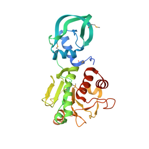
    Purine nucleosides on position 9 of eukaryal and archaeal tRNAs are frequently modified in vivo by the post-transcriptional addition of a methyl group on their N1 atom. The methyltransferase Trm10 is responsible for this modification in both these domains of life. While certain Trm10 orthologues specifically methylate either guanosine or adenosine at position 9 of tRNA, others have a dual specificity. Until now structural information about this enzyme family was only available for the catalytic SPOUT domain of Trm10 proteins that show specificity toward guanosine. Here, we present the first crystal structure of a full length Trm10 orthologue specific for adenosine, revealing next to the catalytic SPOUT domain also N- and C-terminal domains. This structure hence provides crucial insights in the tRNA binding mechanism of this unique monomeric family of SPOUT methyltransferases. Moreover, structural comparison of this adenosine-specific Trm10 orthologue with guanosine-specific Trm10 orthologues suggests that the N1 methylation of adenosine relies on additional catalytic residues.


  • Organizational Affiliation

    Structural Biology Brussels, Vrije Universiteit Brussel, Pleinlaan 2, 1050 Brussel, Belgium Structural Biology Research Center, VIB, Pleinlaan 2, 1050 Brussel, Belgium.


Macromolecules
Find similar proteins by:  (by identity cutoff)  |  3D Structure
Entity ID: 1
MoleculeChains Sequence LengthOrganismDetailsImage
TRNA (ADENINE(9)-N1)-METHYLTRANSFERASE269Sulfolobus acidocaldariusMutation(s): 0 
EC: 2.1.1.218
UniProt
Find proteins for Q4J894 (Sulfolobus acidocaldarius (strain ATCC 33909 / DSM 639 / JCM 8929 / NBRC 15157 / NCIMB 11770))
Explore Q4J894 
Go to UniProtKB:  Q4J894
Entity Groups  
Sequence Clusters30% Identity50% Identity70% Identity90% Identity95% Identity100% Identity
UniProt GroupQ4J894
Sequence Annotations
Expand
  • Reference Sequence
Small Molecules
Ligands 1 Unique
IDChains Name / Formula / InChI Key2D Diagram3D Interactions
PEG
Query on PEG

Download Ideal Coordinates CCD File 
B [auth A]DI(HYDROXYETHYL)ETHER
C4 H10 O3
MTHSVFCYNBDYFN-UHFFFAOYSA-N
Modified Residues  1 Unique
IDChains TypeFormula2D DiagramParent
MSE
Query on MSE
A
L-PEPTIDE LINKINGC5 H11 N O2 SeMET
Experimental Data & Validation

Experimental Data

  • Method: X-RAY DIFFRACTION
  • Resolution: 2.40 Å
  • R-Value Free: 0.250 
  • R-Value Work: 0.179 
  • R-Value Observed: 0.182 
  • Space Group: P 21 21 21
Unit Cell:
Length ( Å )Angle ( ˚ )
a = 41.73α = 90
b = 46.4β = 90
c = 139.04γ = 90
Software Package:
Software NamePurpose
XDSdata reduction
XSCALEdata scaling
SHELXDEphasing
autoSHARPphasing
REFMACrefinement

Structure Validation

View Full Validation Report


Entry History 

Deposition Data

Revision History  (Full details and data files)

  • Version 1.0: 2016-01-13
    Type: Initial release
  • Version 1.1: 2016-02-10
    Changes: Database references
  • Version 1.2: 2017-08-23
    Changes: Data collection, Refinement description