5A71

Open and closed conformations and protonation states of Candida antarctica Lipase B: atomic resolution native


Experimental Data Snapshot

  • Method: X-RAY DIFFRACTION
  • Resolution: 0.91 Å
  • R-Value Free: 0.132 
  • R-Value Work: 0.112 
  • R-Value Observed: 0.113 

wwPDB Validation   3D Report Full Report


This is version 2.1 of the entry. See complete history


Literature

Open and Closed States of Candida Antarctica Lipase B: Protonation and the Mechanism of Interfacial Activation.

Stauch, B.Fisher, S.J.Cianci, M.

(2015) J Lipid Res 56: 2348

  • DOI: https://doi.org/10.1194/jlr.M063388
  • Primary Citation of Related Structures:  
    5A6V, 5A71

  • PubMed Abstract: 

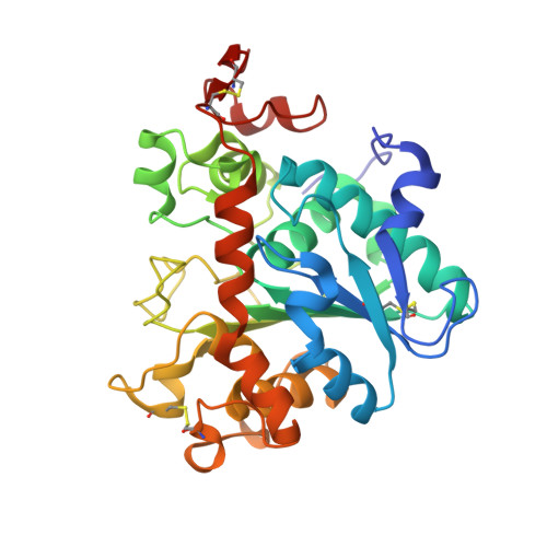
    Lipases (EC 3.1.1.3) are ubiquitous hydrolases for the carboxyl ester bond of water-insoluble substrates, such as triacylglycerols, phospholipids, and other insoluble substrates, acting in aqueous as well as in low-water media, thus being of considerable physiological significance with high interest also for their industrial applications. The hydrolysis reaction follows a two-step mechanism, or "interfacial activation," with adsorption of the enzyme to a heterogeneous interface and subsequent enhancement of the lipolytic activity. Among lipases, Candida antarctica lipase B (CALB) has never shown any significant interfacial activation, and a closed conformation of CALB has never been reported, leading to the conclusion that its behavior was due to the absence of a lid regulating the access to the active site. The lid open and closed conformations and their protonation states are observed in the crystal structure of CALB at 0.91 Å resolution. Having the open and closed states at atomic resolution allows relating protonation to the conformation, indicating the role of Asp145 and Lys290 in the conformation alteration. The findings explain the lack of interfacial activation of CALB and offer new elements to elucidate this mechanism, with the consequent implications for the catalytic properties and classification of lipases.


  • Organizational Affiliation

    European Molecular Biology Laboratory-European Bioinformatics Institute (EMBL-EBI), Wellcome Trust Genome Campus, Hinxton, Cambridge CB10 1SD, United Kingdom Robinson College, University of Cambridge, Cambridge CB3 9AN, United Kingdom.


Macromolecules
Find similar proteins by:  (by identity cutoff)  |  3D Structure
Entity ID: 1
MoleculeChains Sequence LengthOrganismDetailsImage
LIPASE B
A, B
317Moesziomyces antarcticusMutation(s): 0 
EC: 3.1.1.3
UniProt
Find proteins for P41365 (Pseudozyma antarctica)
Explore P41365 
Go to UniProtKB:  P41365
Entity Groups  
Sequence Clusters30% Identity50% Identity70% Identity90% Identity95% Identity100% Identity
UniProt GroupP41365
Sequence Annotations
Expand
  • Reference Sequence
Oligosaccharides

Help

Entity ID: 2
MoleculeChains Length2D Diagram Glycosylation3D Interactions
2-acetamido-2-deoxy-beta-D-glucopyranose-(1-4)-2-acetamido-2-deoxy-beta-D-glucopyranose
C, D
2N-Glycosylation
Glycosylation Resources
GlyTouCan:  G42666HT
GlyCosmos:  G42666HT
GlyGen:  G42666HT
Small Molecules
Ligands 3 Unique
IDChains Name / Formula / InChI Key2D Diagram3D Interactions
IPA
Query on IPA

Download Ideal Coordinates CCD File 
E [auth A],
J [auth B],
K [auth B]
ISOPROPYL ALCOHOL
C3 H8 O
KFZMGEQAYNKOFK-UHFFFAOYSA-N
K
Query on K

Download Ideal Coordinates CCD File 
F [auth A],
G [auth A],
H [auth A],
L [auth B]
POTASSIUM ION
K
NPYPAHLBTDXSSS-UHFFFAOYSA-N
NA
Query on NA

Download Ideal Coordinates CCD File 
I [auth A],
M [auth B],
N [auth B],
O [auth B],
P [auth B]
SODIUM ION
Na
FKNQFGJONOIPTF-UHFFFAOYSA-N
Experimental Data & Validation

Experimental Data

  • Method: X-RAY DIFFRACTION
  • Resolution: 0.91 Å
  • R-Value Free: 0.132 
  • R-Value Work: 0.112 
  • R-Value Observed: 0.113 
  • Space Group: P 1
Unit Cell:
Length ( Å )Angle ( ˚ )
a = 39.67α = 88.74
b = 48.94β = 97.15
c = 71.61γ = 108.44
Software Package:
Software NamePurpose
REFMACrefinement
XDSdata reduction
Aimlessdata scaling
MOLREPphasing

Structure Validation

View Full Validation Report


Entry History 

Deposition Data

Revision History  (Full details and data files)

  • Version 1.0: 2015-10-21
    Type: Initial release
  • Version 1.1: 2015-12-16
    Changes: Database references
  • Version 1.2: 2018-03-07
    Changes: Data collection, Source and taxonomy
  • Version 1.3: 2019-03-06
    Changes: Data collection, Derived calculations, Experimental preparation
  • Version 1.4: 2019-05-15
    Changes: Data collection, Experimental preparation
  • Version 2.0: 2020-07-29
    Type: Remediation
    Reason: Carbohydrate remediation
    Changes: Atomic model, Data collection, Derived calculations, Other, Structure summary
  • Version 2.1: 2024-01-10
    Changes: Data collection, Database references, Refinement description, Structure summary