5A5F

CRYSTAL STRUCTURE OF MURD LIGASE FROM ESCHERICHIA COLI IN COMPLEX WITH UMA AND ADP


Experimental Data Snapshot

  • Method: X-RAY DIFFRACTION
  • Resolution: 1.90 Å
  • R-Value Free: 0.232 
  • R-Value Work: 0.198 
  • R-Value Observed: 0.200 

wwPDB Validation   3D Report Full Report


Ligand Structure Quality Assessment 


This is version 1.2 of the entry. See complete history


Literature

Crystallographic Study of Peptidoglycan Biosynthesis Enzyme MurD: Domain Movement Revisited.

Sink, R.Kotnik, M.Zega, A.Barreteau, H.Gobec, S.Blanot, D.Dessen, A.Contreras-Martel, C.

(2016) PLoS One 11: e0152075-e0152075

  • DOI: https://doi.org/10.1371/journal.pone.0152075
  • Primary Citation of Related Structures:  
    5A5E, 5A5F

  • PubMed Abstract: 

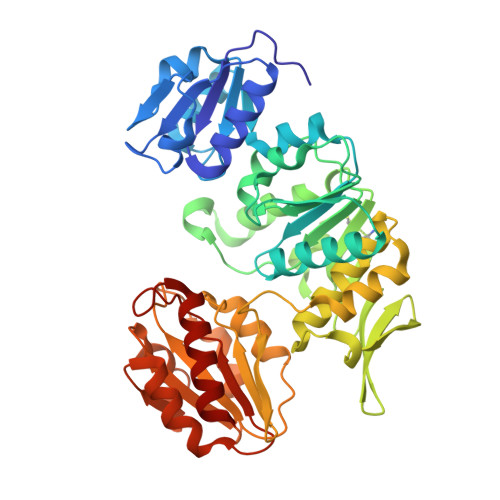
    The biosynthetic pathway of peptidoglycan, an essential component of bacterial cell wall, is a well-recognized target for antibiotic development. Peptidoglycan precursors are synthesized in the bacterial cytosol by various enzymes including the ATP-hydrolyzing Mur ligases, which catalyze the stepwise addition of amino acids to a UDP-MurNAc precursor to yield UDP-MurNAc-pentapeptide. MurD catalyzes the addition of D-glutamic acid to UDP-MurNAc-L-Ala in the presence of ATP; structural and biochemical studies have suggested the binding of the substrates with an ordered kinetic mechanism in which ligand binding inevitably closes the active site. In this work, we challenge this assumption by reporting the crystal structures of intermediate forms of MurD either in the absence of ligands or in the presence of small molecules. A detailed analysis provides insight into the events that lead to the closure of MurD and reveals that minor structural modifications contribute to major overall conformation alterations. These novel insights will be instrumental in the development of new potential antibiotics designed to target the peptidoglycan biosynthetic pathway.


  • Organizational Affiliation

    University of Ljubljana, Faculty of Pharmacy, Aškerčeva 7, Ljubljana, Slovenia.


Macromolecules
Find similar proteins by:  (by identity cutoff)  |  3D Structure
Entity ID: 1
MoleculeChains Sequence LengthOrganismDetailsImage
UDP-N-ACETYLMURAMOYLALANINE--D-GLUTAMATE LIGASE438Escherichia coli K-12Mutation(s): 0 
EC: 6.3.2.9
UniProt
Find proteins for P14900 (Escherichia coli (strain K12))
Explore P14900 
Go to UniProtKB:  P14900
Entity Groups  
Sequence Clusters30% Identity50% Identity70% Identity90% Identity95% Identity100% Identity
UniProt GroupP14900
Sequence Annotations
Expand
  • Reference Sequence
Experimental Data & Validation

Experimental Data

  • Method: X-RAY DIFFRACTION
  • Resolution: 1.90 Å
  • R-Value Free: 0.232 
  • R-Value Work: 0.198 
  • R-Value Observed: 0.200 
  • Space Group: P 21 21 21
Unit Cell:
Length ( Å )Angle ( ˚ )
a = 66.44α = 90
b = 89.84β = 90
c = 108.54γ = 90
Software Package:
Software NamePurpose
REFMACrefinement
XDSdata reduction
XSCALEdata scaling
PHASERphasing

Structure Validation

View Full Validation Report


Ligand Structure Quality Assessment 


Entry History 

Deposition Data

Revision History  (Full details and data files)

  • Version 1.0: 2016-04-13
    Type: Initial release
  • Version 1.1: 2018-02-28
    Changes: Database references, Source and taxonomy
  • Version 1.2: 2024-01-10
    Changes: Data collection, Database references, Derived calculations, Other, Refinement description