5A3S

Crystal structure of the (SR) Calcium ATPase E2-vanadate complex bound to thapsigargin and TNP-ATP


Experimental Data Snapshot

  • Method: X-RAY DIFFRACTION
  • Resolution: 3.30 Å
  • R-Value Free: 0.245 
  • R-Value Work: 0.210 
  • R-Value Observed: 0.211 

wwPDB Validation   3D Report Full Report


Ligand Structure Quality Assessment 


This is version 1.4 of the entry. See complete history


Literature

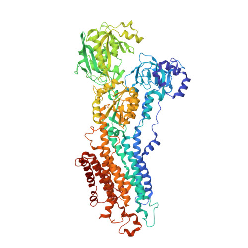
Crystal Structure of the Vanadate-Inhibited Ca(2+)-ATPase.

Clausen, J.D.Bublitz, M.Arnou, B.Olesen, C.Andersen, J.P.Moller, J.V.Nissen, P.

(2016) Structure 24: 617

  • DOI: https://doi.org/10.1016/j.str.2016.02.018
  • Primary Citation of Related Structures:  
    5A3Q, 5A3R, 5A3S

  • PubMed Abstract: 

    Vanadate is the hallmark inhibitor of the P-type ATPase family; however, structural details of its inhibitory mechanism have remained unresolved. We have determined the crystal structure of sarcoplasmic reticulum Ca(2+)-ATPase with bound vanadate in the absence of Ca(2+). Vanadate is bound at the catalytic site as a planar VO3(-) in complex with water and Mg(2+) in a dephosphorylation transition-state-like conformation. Validating bound VO3(-) by anomalous difference Fourier maps using long-wavelength data we also identify a hitherto undescribed Cl(-) site near the dephosphorylation site. Crystallization was facilitated by trinitrophenyl (TNP)-derivatized nucleotides that bind with the TNP moiety occupying the binding pocket that normally accommodates the adenine of ATP, rationalizing their remarkably high affinity for E2P-like conformations of the Ca(2+)-ATPase. A comparison of the configurations of bound nucleotide analogs in the E2·VO3(-) structure with that in E2·BeF3(-) (E2P ground state analog) reveals multiple binding modes to the Ca(2+)-ATPase.


  • Organizational Affiliation

    Department of Molecular Biology and Genetics, Aarhus University, 8000 Aarhus, Denmark; Centre for Membrane Pumps in Cells and Disease - PUMPKIN, Danish National Research Foundation, Aarhus University, 8000 Aarhus, Denmark; Department of Biomedicine, Aarhus University, 8000 Aarhus, Denmark.


Macromolecules
Find similar proteins by:  (by identity cutoff)  |  3D Structure
Entity ID: 1
MoleculeChains Sequence LengthOrganismDetailsImage
SARCOPLASMIC RETICULUM CALCIUM ATPASE 1 MOLECULE SARCOPLASMIC/ENDOPLASMIC RETICULUM CALCIUM ATPASE 1
A, B
995Oryctolagus cuniculusMutation(s): 1 
EC: 3.6.3.8
Membrane Entity: Yes 
UniProt
Find proteins for P04191 (Oryctolagus cuniculus)
Explore P04191 
Go to UniProtKB:  P04191
Entity Groups  
Sequence Clusters30% Identity50% Identity70% Identity90% Identity95% Identity100% Identity
UniProt GroupP04191
Sequence Annotations
Expand
  • Reference Sequence
Small Molecules
Ligands 6 Unique
IDChains Name / Formula / InChI Key2D Diagram3D Interactions
128
Query on 128

Download Ideal Coordinates CCD File 
E [auth A],
L [auth B]
SPIRO(2,4,6-TRINITROBENZENE[1,2A]-2O',3O'-METHYLENE-ADENINE-TRIPHOSPHATE
C16 H17 N8 O19 P3
XFMMHXLUHKBKQE-UHEGPQQHSA-N
TG1
Query on TG1

Download Ideal Coordinates CCD File 
C [auth A],
J [auth B]
OCTANOIC ACID [3S-[3ALPHA, 3ABETA, 4ALPHA, 6BETA, 6ABETA, 7BETA, 8ALPHA(Z), 9BALPHA]]-6-(ACETYLOXY)-2,3,-3A,4,5,6,6A,7,8,9B-DECAHYDRO-3,3A-DIHYDROXY-3,6,9-TRIMETHYL-8-[(2-METHYL-1-OXO-2-BUTENYL)OX Y]-2-OXO-4-(1-OXOBUTOXY)-AZULENO[4,5-B]FURAN-7-YL ESTER
C34 H50 O12
IXFPJGBNCFXKPI-FSIHEZPISA-N
VN4
Query on VN4

Download Ideal Coordinates CCD File 
D [auth A],
K [auth B]
oxido(dioxo)vanadium
O3 V
ALTWGIIQPLQAAM-UHFFFAOYSA-N
K
Query on K

Download Ideal Coordinates CCD File 
H [auth A],
O [auth B]
POTASSIUM ION
K
NPYPAHLBTDXSSS-UHFFFAOYSA-N
CL
Query on CL

Download Ideal Coordinates CCD File 
I [auth A],
P [auth B]
CHLORIDE ION
Cl
VEXZGXHMUGYJMC-UHFFFAOYSA-M
MG
Query on MG

Download Ideal Coordinates CCD File 
F [auth A],
G [auth A],
M [auth B],
N [auth B]
MAGNESIUM ION
Mg
JLVVSXFLKOJNIY-UHFFFAOYSA-N
Binding Affinity Annotations 
IDSourceBinding Affinity
TG1 BindingDB:  5A3S Ki: 0.1 (nM) from 1 assay(s)
Kd: 0.2 (nM) from 1 assay(s)
IC50: min: 0.2, max: 3.9 (nM) from 2 assay(s)
Experimental Data & Validation

Experimental Data

  • Method: X-RAY DIFFRACTION
  • Resolution: 3.30 Å
  • R-Value Free: 0.245 
  • R-Value Work: 0.210 
  • R-Value Observed: 0.211 
  • Space Group: P 1 21 1
Unit Cell:
Length ( Å )Angle ( ˚ )
a = 130.556α = 90
b = 93.778β = 107.26
c = 135.692γ = 90
Software Package:
Software NamePurpose
PHENIXrefinement
XDSdata reduction
XSCALEdata scaling
PHASERphasing

Structure Validation

View Full Validation Report


Ligand Structure Quality Assessment 


Entry History 

Deposition Data

Revision History  (Full details and data files)

  • Version 1.0: 2016-04-13
    Type: Initial release
  • Version 1.1: 2016-04-20
    Changes: Database references
  • Version 1.2: 2019-05-15
    Changes: Data collection, Derived calculations, Experimental preparation, Other
  • Version 1.3: 2019-10-23
    Changes: Data collection, Database references, Other
  • Version 1.4: 2024-01-10
    Changes: Data collection, Database references, Derived calculations, Refinement description, Structure summary